Laser-Doppler-Bildgebung

#moorLDI2-IR

Grossflächige Laser-Doppler-Bildgebung mit hoher Auflösung

  • I cannot rate the company or the staff highly enough.

    Jim House, PhD
    University of Portsmouth

  • We can't recommend Moor instruments highly enough. The technology is at the cutting edge and the support second to none.

    Paul Sumners, PhD
    London South Bank University

  • We have found Moor equipment to be extremely dependable and innovative.

    Dean L. Kellogg, Jr., MD, Ph.D
    University of Texas Health Science Center

  • Laser Doppler Imager is a standard accurate method we now use in our cerebral blood flow and brain perfusion in our laboratory.

    Momoh A. Yakubu, PhD
    Texas Southern University

  • Moor Instruments have consistently provided excellent help and support for my research.

    Kim Gooding, PhD
    University of Exeter Medical School

  • It goes without saying that the company's imaging technology itself is superb!

    Gourav Banerjee
    Leeds Beckett University

  • In a nutshell, moorFLPI-2 is the most user-friendly system for studying cerebral blood flow regulation in rodents.

    Chia-Yi (Alex) Kuan, MD, PhD
    Emory University School of Medicine

  • I expect to be using Moor Instrument’s technology for many years to come!

    Faisel Khan, PhD
    Ninewells Hospital & Medical School

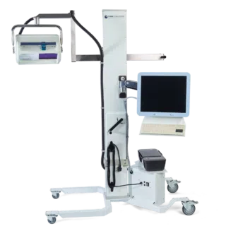
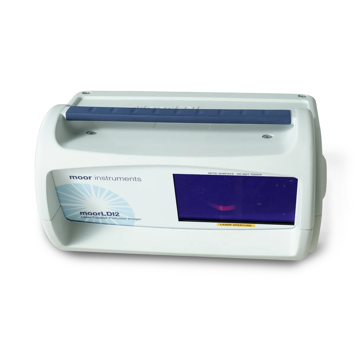
Die moorLDI2-IR Laser-Doppler-Imager bieten eine gut etablierte, hochwertige Lösung zur Darstellung des Blutflusses im Gewebe für Ihre Anwendungen in der Klinik oder in der Grundlagenforschung. Das System wird routinemäßig in zahlreichen Laboratorien und Kliniken weltweit eingesetzt und vereint ein einzigartiges optisches Design mit spezieller Signalverarbeitung, um die höchste Auflösung und die schärfsten Bilder in dieser Produktklasse zu liefern.

Laser-Doppler-Imaging (LDI) wird oft mit dem Laser-Speckle-Imaging verglichen und obwohl es einige Gemeinsamkeiten gibt, bieten beide Techniken eigene Vorteile. LDI bietet generell eine höhere Eindringtiefe, die die Visualisierung von kleinen Gefäßen unter der Gewebeoberfläche erlaubt. Dies ist optimal für die Darstellung der Angiogenese oder für Messungen des cerebralen Blutflusses durch die Schädeldecke.

Der moorLDI2-IR erlaubt ebenfalls die Darstellung einer sehr großen Fläche – bis zu 50 cm x 50 cm in einem Scan. Für einige Anwendungen ist dies eine notwendige Bedingung.

Andere Funktionen und Vorteile sind:

  • Berührungslose Messungen – schmerzfrei für Patienten, keine Infektionsgefahr, keine chemischen Kontrastmittel oder Farben nötig.
  • Verwendung bei Raumbeleuchtung – in den meisten wissenschaftlichen, klinischen Räumlichkeiten und im OP-Bereich.
  • Flexible Scangröße – von der Größe einer Fingerspitze bis zur Größe eines Oberkörpers.
  • Hohe räumliche Auflösung – um die kleinsten Details bis zu 100 µm darzustellen.
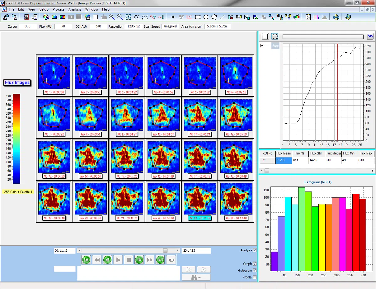
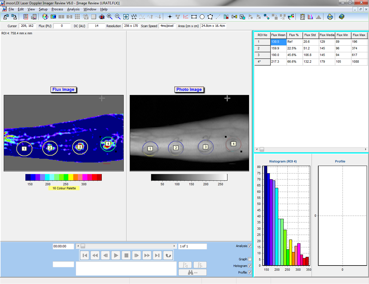
  • Einzelbild und automatisierte Bildgebung – Vergleich des Blutflusses von Region zu Region innerhalb desselben Scans und wiederholtes Scannen derselben Region über längere Zeit.
  • Fortschrittliche Windows-kompatible Software – diese erleichtert die Einstellungen und das Scannen. Vielseitige Analysefunktionen ermöglichen die bestmögliche Verarbeitung der erfassten Daten.
  • Protokollkontrolle – Programmierung von Hitzestimulation, Druckmanschetten und transdermaler Medikamentengabe. Reproduzierbar, präzise und verlässlich.
  • Digitale Trigger für Ein- und Ausgänge – zur Synchronisierung mit externen Geräten.
  • Digitale Signalverarbeitung und hochqualitative Optik bieten höchste Sensitivität bei Veränderungen des Blutflusses und hervorragende Zuverlässigkeit.
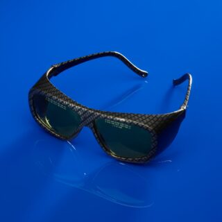
  • Auswahl der Laserwellenlänge – zur Messung von oberflächlichen oder etwas tieferen Gewebeschichten.
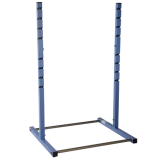
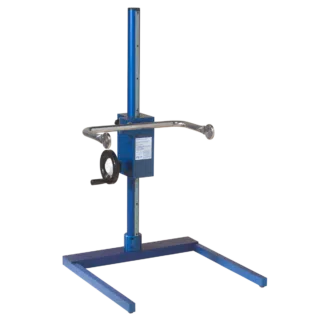
  • Auswahl der Befestigung – von Tischhalterungen bis zu Rollwagen für die Verwendung im OP.

Das folgende Zubehör für den moorLDI2-IR können Sie ONLINE KAUFEN


Der nachfolgende Abschnitt enthält eine Liste der häufigsten Fragen unserer Kunden. Falls Sie eine Frage haben und Sie die Antwort darauf hier nicht finden, senden Sie uns bitte eine Email. Wir helfen Ihnen gerne!


Q. Was ist der größte Bildbereich in einem Perfusionsbild?
A. Alle Varianten des moorLDI2 mit Ausnahme des HIR (high resolution)-Modells scannen ein Areal von bis zu 50 cm x 50 cm Größe. Um eine hohe räumliche Auflösung zu erreichen kann der moorLDI2-HIR verwendet werden, der 2,5 cm x 2,5 cm mit 256 x 256 Pixel erfasst – ungefähr 100 Pixel pro mm2.

Q. Wie oft sollte der moorLDI2 kalibriert werden?
A. Mit dem System liefern wir einen Kalibrationsblock, mit dem der Benutzer die Kalibration selbst einfach testen und durchführen kann. Wir empfehlen einen wöchentlichen Test, eine Kalibration ist aber nur selten erforderlich.

Q. Wie kann ich mit dem moorLDI2-IR Perfusionsantworten auf Iontophorese oder Hauterwärmung testen?
A. Dazu kann der Imager so eingestellt werden, dass eine definierte Anzahl von Perfusionsbildern in Folge mit definierte Zwischenzeiten gescannt werden. Transparente Erwärmungs- und Iontophoresekammern sind von uns erhältlich, die die Beobachtung des stimulierten Areals und des Kontrollgewebes ermöglichen.

Q. Was ist der Vorteil eines kontinuierlich scannenden Laserstrahls?
A. Der moorLDI2 verwendet ein patentiertes Verfahren zum kontinuierlichen Scannen des Laserstrahls, d.h. der Laser wird zwischen den einzelnen Messpunkten nicht abgeschaltet und ermöglicht dadurch eine deutlich höhere Scangeschwindigkeit. Die erreichte Scangeschwindigkeit und eine hohe Bildrate sind besonders für klinische Anwendungen wichtig. Für Anwendungen, in denen besonders kleine Perfusionsraten erfasst werden müssen, empfehlen wir eine Herabsetzung der Scanrate durch die Software.

There are numerous references where our laser Doppler imagers are cited. The list below is a small selection. Please contact us for reference lists on your chosen subject.


Abraham, A., Alabdali, M., Alsulaiman, A., Breiner, A., Barnett, C., Katzberg, H.D., Lovblom, L.E., Perkins, B.A., Bril, V., (2016).
Laser Doppler Flare Imaging and Quantitative Thermal Thresholds Testing Performance in Small and Mixed Fiber Neuropathies.
Plos One, 11(11), ppe0165731.

Campos-Junior, P. H. A., Alves, T. J. M., Dias, M. T., Assunçao, C. M., Munk, M., Mattos, M. S., Kraemer, L. R., Almeida, B. G., Russo, R. C., Barcelos, L., Camargo, L. S. A., and Viana, J. H. M., (2016).
Ovarian Grafts 10 Days after Xenotransplantation: Folliculogenesis and Recovery of Viable Oocytes.
PLoS ONE 11(6): e0158109. doi:10.1371/journal.pone.0158109.

Chalothorn, D., Clayton, J .A, Zhang, H., Pomp , D., and Faber, J. E., (2007).
Collateral density, remodeling, and VEGF-A expression differ widely between mouse strains.
Physiological genomics, 30(2), pp.179–91.

Datta , D., Ferrell, W . R., Sturrock , R . D., Jadhav , S . T., and Sattar , N., (2007).
Inflammatory suppression rapidly attenuates microvascular dysfunction in rheumatoid arthritis.
Atherosclerosis, 192(2), pp.391–5.

De, P., Peng , Q., Traktuevc, D . O., Li , W., Yoder, M . C., March, K . L., Durden, D . L. (2009).
Expression of RAC2 in endothelial cells is required for the postnatal neovascular response.
Experimental Cell Research, 315(2), pp.248–263.

Dimitroulas, T., Sandoo, A., Hodson, J., Smith, J. P., and Kitas, G. D., (2016).
In vivo microvascular and macrovascular endothelial function is not associated with circulating dimethylarginines in patients with rheumatoid arthritis: a prospective analysis of the DRACCO cohort.
Scand J Clin Lab Invest, 76(4), pp:331-7.

Ebadi, H., Perkins, B . a, Katzberg, H . D., Lovblom, L . E., Bril, V.,(2012).
Evaluation of proxy tests for SFSN: evidence for mixed small and large fiber dysfunction.
PloS one, 7(8), p.e42208.

Edmunds, MC., Wigmore, S., Kluth, D., (2013).
In situ transverse rectus abdominis myocutaneous flap: a rat model of myocutaneous ischemia reperfusion injury.
J Vis Exp. 2013 Jun 8;(76). www.jove.com/video/50473/in-situ-transverse-rectus-abdominis-myocutaneous-flap-rat-model

Gil, C. H., Ki, B. S., Seo, J., Choi, J. J., Kim, H., Kim, I. G., Jung, A. R., Lee, W-Y., Choi, Y., Park, K., Moon, S-H., and Chung, H-M., (2016).
Directing human embryonic stem cells towards functional endothelial cells easily and without purification.
Tissue Engineering and Regenerative Medicine. 13(3), pp:274–283.

Gorodkin, R., Herrick, A. L., and Murray, A. K., (2016).
Microvascular response in patients with complex regional pain syndrome as measured by laser Doppler imaging.
Microcirculation, 23(5), pp:379-83.

Gunawardena, H., Harris, N . D., Carmichael, C., McHugh, N . J., (2007a).
Maximum blood flow and microvascular regulatory responses in systemic sclerosis.
Rheumatology (Oxford, England), 46(7), pp.1079–82.

Gunawardena, H., Harris, N . D., Carmichael, C., McHugh, N . J., (2007b).
Microvascular responses following digital thermal hyperaemia and iontophoresis measured by laser Doppler imaging in idiopathic inflammatory myopathy.
Rheumatology (Oxford, England), 46(9), pp.1483–6.

Hsieh, M.J., Liu, H.T., Wang, C.N., Huang, H.Y., Lin, Y., Ko, Y.S., Wang, J.S., Chang, V.H., and Pang, J.S., (2016).
Therapeutic potential of pro-angiogenic BPC157 is associated with VEGFR2 activation and up-regulation.
J Mol Med (Berl), Nov 15.[Epub ahead of print].

Isenberg, J . S., Maxhimer , J . B., Powers, P., Tsokos, M ., Frazier, W . a, Roberts , D . D., (2008).
Treatment of liver ischemia-reperfusion injury by limiting thrombospondin-1/CD47 signaling.
Surgery, 144(5), pp.752–61.

Isenberg , J . S., Shiva, S., Gladwin, M., (2009).
Thrombospondin-1-CD47 blockade and exogenous nitrite enhance ischemic tissue survival, blood flow and angiogenesis via coupled NO-cGMP pathway activation.
Nitric oxide : biology and chemistry / official journal of the Nitric Oxide Society, 21(1), pp.52–62.

Khoo, T . L., Halim, a S., Zakaria, Z., Mat Saad, a Z., Wu, L . Y., Lau, H . Y.,(2011).
A prospective, randomised, double-blinded trial to study the efficacy of topical tocotrienol in the prevention of hypertrophic scars. Journal of plastic, reconstructive & aesthetic surgery :
JPRAS, 64(6), pp.e137–45.

Krishnan, S . T., Quattrini, C., Jeziorska M., Malik, R.A., Rayman, G., (2007).
Neurovascular Factors in Wound Healing in the Foot Skin of Type 2 Diabetic Subjects.
DIABETES CARE,, 30(12), pp.3058 – 3062.

Kvietys, P., Granger, D., (2012).
Role of reactive oxygen and nitrogen species in the vascular responses to inflammation.
Free Radical Biology and Medicine, 52(3), pp.556–592.

Jensen, A. R., Manning, M. M., Khaneki, S., Drucker, N. A., and Markel, T. A., (2016).
Harvest Tissue Source Does Not Alter the Protective Power of Stromal Cell Therapy Following Intestinal Ischemia and Reperfusion Injury.
Journal of SurgicalResearch, 204(2), pp:361-370.

Lasch, M., Caballero-Martinez, A., Troidl, K., Schloegl, I., Lautz, T., and Deindl, E., (2016).
Arginase inhibition attenuates arteriogenesis and interferes with M2 macrophage accumulation.
Lab Invest. 2016 May 30, doi: 10.1038/labinvest.2016.62. [Epub ahead of print].

Lee, J. H., Ryu, J. M., Han, Y. S., Zia, M. F., Kwon, H. Y., Noh, H., Han, H. J., and Lee, S. H., (2016).
Fucoidan improves bioactivity and vasculogenic potential of mesenchymal stem cells in murine hind limb ischemia associated with chronic kidney disease.
J Mol Cell Cardiol, 97, pp:169-17.

Lohman , B., (2007).
The effect of whole body vibration on lower extremity skin blood flow in normal subjects.
Med Sci …, 13(2).

Marks, D. J. B., Harbord, M . W. N., Macallister, R., Rahman, F . Z., Young , J., Al-lazikani, B., Lees, W., Novelli, M., Bloom, S., Segal, A . W., (2006).
Defective acute inflammation in Crohn’s disease : a clinical investigation.
Lancet, 367.

Nabavi Nouri, M., Ahmed, A., Bril, V., Orszag, A., Ng, E., Nwe, P., Perkins, B . a, (2012).
Diabetic neuropathy and axon reflex-mediated neurogenic vasodilatation in type 1 diabetes.
PloS one, 7(4), p.e34807.

Oda, M., Toba, K., Ozawa, T., Kato, K., Yanagawa, T., Ikarashi, N., Takayama, T., Suzuki, T., Hanawa, H., Fuse, I., Nakata, K., Narita, M., Takahashi, M., Aizawa, Y., (2010).
Establishment of culturing system for ex-vivo expansion of angiogenic immature erythroid cells, and its application for treatment of patients with chronic severe lower limb ischemia.
Journal of molecular and cellular cardiology, 49(3), pp.347–53.

Pinto, N.C., Cassini-Vieira, P., de Souza-Fagundes, E.M., Barcelos, L.S., Nogueira Castañon, M.C., and Scio, E., 2016.
Pereskia aculeata Miller leaves accelerate excisional wound healing in mice.
J Ethnopharmacol., Sep 3. [Epub ahead of print].

Silva, L.P., Pirraco, R.P., Santos, T.C., Novoa-Carballal, R., Cerqueira, M.T.,2, Reis R.L., Correlo, V.M., and Marques, A.P., (2016).
Neovascularization Induced by the Hyaluronic Acid-Based Spongy-Like Hydrogels Degradation Products.
ACS Appl Mater Interfaces, 8(49). pp33464-33474.

Terkelsen, A. J., Bach, F. W., Jensen, T . S., (2009).
Experimental forearm immobilization in humans reduces capsaicin-induced pain and flare.
Brain research, 1263, pp.43–9.

Tsatralis, T., Ridiandries, A., Robertson, S., Vanags, L.Z., Lam, Y.T., Tan, J.T.M., Ng, M.K.C., and Bursill, C.A., 2016.
Reconstituted high-density lipoproteins promote wound repair and blood flow recovery in response to ischemia in aged mice.
Lipids in Health and Disease . 15(1), pp150-161.

Vasam, G., Joshi, S., Jarajapu, Y. P. R., (2016).
Impaired Mobilization of Vascular Reparative Bone Marrow Cells in Streptozotocin-Induced Diabetes but not in Leptin Receptor-Deficient db/db Mice.
Scientific Reports 6(26131), pp:1-13.

Wajima, D., Nakamura, M., Horiuchi, K., Miyake, H., Takeshima, Y., Tamura, K., Motoyama, Y., Konishi, N., Nakase, H., (2010).
Enhanced cerebral ischemic lesions after two-vein occlusion in diabetic rats.
Brain research, 1309, pp.126–35.

Wester, T., Häggblad, E., (2011).
Assessments of skin and tongue microcirculation reveals major changes in porcine sepsis.
Clinical Physiology

Winchester, L., Chou,N ., (2008).
Measurement of sublingual blood velocity as a tool for monitoring sepsis.
Engineering in Medicine and …, (1), pp.3739–3742.

Yang, Y., Chen, Z., Zhang, T., Wang, S., and Qing, Y., (2016).
Wnt3a promoted the therapeutic of angiogenesis on lower leg ischemia with endothelial progenitor cells.
Int J Clin Exp Med, 9(3), pp:5902-5911.

Zeng, L., Xiao, Q., Chen, M., Margariti, A., Martin, D., Ivetic, A., Xu, H., Mason, J., Wang, W., Cockerill, G., Mori, K., Li, J . Y., Chien, S., Hu, Y., Xu, Q., (2013).
Vascular endothelial cell growth-activated XBP1 splicing in endothelial cells is crucial for angiogenesis.
Circulation, 127(16), pp.1712–22.

Zhao, Y., Li, Y., Luo, P., Gao, Y., Yang, J., Lao, K-H., Wang, G., Cockerill, G., Hu, Y., Xu, Q., Li, T., and Zeng, L., (2016).
XBP1 splicing triggers miR-150 transfer from smooth muscle cells to endothelial cells via extracellular vesicles.
Sci. Rep. 6, 28627.

Moor Instruments are committed to product development. We reserve the right to change the specifications below without notice.

The moorLDI2-IR is a Class IIa Active device for diagnosis under EC directive 93/42/EEC 14th June 1993 Medical Device Directive.


LASER SOURCE

Infrarot-Laserdiode: 785 nm nominal , maximale Leistung 2,5 mW
Entfernung für Augengefahr: 20 m.
Klasse 3R nach IEC 60825-1:2014 und entspricht den FDA Performancestandards für Laserprodukte, ausgenommen Abweichungen entsprechend Laserankündigung No. 50 vom 24. Juni, 2007.
Sichtbare Laserdiode (Zielstrahl bei Infrarotgeräten): 660 nm nominal, maximale Leistung 0,25 mW.
Alle Angaben beinhalten kumulative Messungenauigkeiten und mögliche Zunahmen nach Herstellung.

AUGENSCHUTZMASSNAHMEN

Die nominelle Augenschutzentfernung beträgt 20 m.
Anwenderschutz OD4, 770-850 nm
Patientenschutz OD4, 630-670 nm und 770-850 nm

BETRIEBSBEDINGUNGEN

Temperatur: 15°C bis 30°C
Luftfeuchtigkeit: 45% bis 75%
Luftdruck: innerhalb von 86,0 kPa bis 106,0 kPa (entsprechend 645 mmHg bis 795 mmHg).
Entflammbare Anästhetika: Das System darf nicht bei Anwesenheit von entflammbaren Anästhetika verwendet werden.

BANDBREITE

Abhängig von der Scanrate: Tiefpass (3db): 20 Hz, 100 Hz oder 250 Hz.
Hochpass wählbar (0.1db): 3 kHz, 15 kHz oder 22,5 kHz.

(Standardeinstellungen in fett).

BILDAREAL

Bei einer Entfernung von 20 cm: Normalbereich = 6,6 cm x 6,6 cm; Großer Bereich = 13 cm x 13 cm.
Bei einer Entfernung von 100 cm: Normalbereich = 25 cm x 25 cm; Großer Bereich = 50 cm x 50 cm.

SCAN-GESCHWINDIGKEIT

Scangeschwindigkeit is ungefähr 4 ms / Pixel, 10 ms / Pixel oder 50 ms / Pixel (bei maixmaler Auflösung).
Scandauer ist typischerweise 40 s für ein 12,5 cm x 12,5 cm Bild mit 64 x 64 Pixel Auflösung, ungefähr 6 min für ein 50 cm x 50 cm Bild mit 256 x 256 Pixel Auflösung bei 4 ms / Pixel und 100 cm Abstand.

RÄUMLICHE AUFLÖSUNG

Bis zu 256 x 256 Pixel (tatsächliche Messwerte und nicht interpoliert): 0,2 mm/Pixel bei 20 cm, „normaler Scan“; 2,0 mm / Pixel bei 100 cm, „großer Scan“.

UMGEBUNGSLICHT

Normales Raumlicht.

MESSWERTE

Fluss (FLUX) – Genauigkeit: ± 10% relative zum Moor Instruments moorLDI2 Standard
Präzision: ± 3% der Messwerte
Messbereich: 0 – 5000 PU (perfusion unit)

Konzentration (CONC) – Genauigkeit: ± 10%
Präzision: ± 5 % der Messwerte
Messbereich: 0 – 5000 w.E. (willkürliche Einheit)

DC Genauigkeit: ± 10%
Präzision: ± 3%
Messbereich: 0 – 5000 w.E.

CCD KAMERA

Autofokus, Motorbetriebener 10 x Zoom. 752 x 582 Pixel-Auflösung.

SOFTWARE

Windows basierte Software für Instrumentensteuerung, Datenerfassung und Auswertung.

ELEKTRISCHE SICHERHEITS KLASSIFIZIERUNG

Sicherheit gegen elektrischen Schlag – Class I.
Sicherheitsgrad gegen elektrischen Schlag – Typ B.
Sicherheitsgrad gegen Eindringen von Flüssigkeiten – IPXO (nicht geschützt).
Schutz gegen entflammbare Anästhetika: Gerät nicht geeignet für den Betrieb bei Anwesenheit von entflammbaren Anästhetika mit Luft oder NO.
Betriebsart: kontinuierlich.

ALLGEMEIN

Universelles Schaltnetzteil.
Spannungsversorgung 84 bis 264 V AC 50 VA, 50 bis 60 Hz.
Kontroll-Einheit: Abmessungen B H T in mm 305 x 115 x 260; Gewicht 4,5 kg.
Scan-Kopf: Abmessungen B H T in mm 426 x 244 x 300; Gewicht 8 kg.
Betriebstemperatur: 15 – 30°C.

ARBEITS- UND TRANSPORTBEDINGUNGEN

Temperatur: 0 – 45°C.
Feuchtigkeit: 0 – 80%.
Luftdruck: 500 hPa – 1060 hPa.