Laser Doppler Imager

#moorLDI2-IR

Large area, high resolution blood flow laser Doppler imaging

  • Laser Doppler Imager is a standard accurate method we now use in our cerebral blood flow and brain perfusion in our laboratory.

    Momoh A. Yakubu, PhD
    Texas Southern University

  • I expect to be using Moor Instrument’s technology for many years to come!

    Faisel Khan, PhD
    Ninewells Hospital & Medical School

  • We have found Moor equipment to be extremely dependable and innovative.

    Dean L. Kellogg, Jr., MD, Ph.D
    University of Texas Health Science Center

  • Moor Instruments have consistently provided excellent help and support for my research.

    Kim Gooding, PhD
    University of Exeter Medical School

  • We can't recommend Moor instruments highly enough. The technology is at the cutting edge and the support second to none.

    Paul Sumners, PhD
    London South Bank University

  • In a nutshell, moorFLPI-2 is the most user-friendly system for studying cerebral blood flow regulation in rodents.

    Chia-Yi (Alex) Kuan, MD, PhD
    Emory University School of Medicine

  • I cannot rate the company or the staff highly enough.

    Jim House, PhD
    University of Portsmouth

  • It goes without saying that the company's imaging technology itself is superb!

    Gourav Banerjee
    Leeds Beckett University

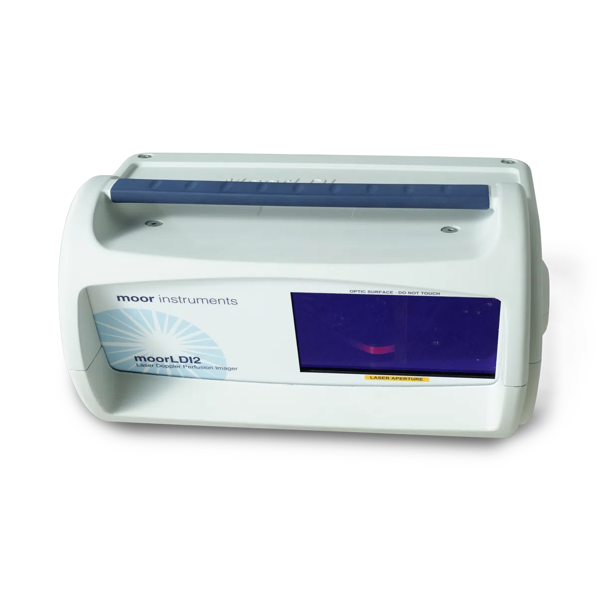
The moorLDI2-IR laser Doppler blood flow imager offers a well proven, high specification solution to your blood flow application for clinical or research application. The system is in routine use in numerous laboratories and clinics globally and employs unique, optical design and signal processing in order to generate the highest resolution and clearest images of its class.

Laser Doppler imaging (LDI) is often compared to laser speckle imaging and whilst there are some similarities, both techniques offer unique advantages. LDI generally offers deeper penetration enabling enhanced visualisation of small vessels below the tissue surface, perfect for angiogenesis modelling or through skull pre clinical cerebral blood flow imaging.

The moorLDI2-IR also allows very large area imaging – up to 50cm x 50cm in one scan. For certain applications these features are critical.

Other features and benefits include;

  • Non contact measurement – painless for patient, aids infection control, no chemical tracers or dyes needed.
  • Daylight operation – use in most lab, clinic or theatre settings.
  • Flexible scan sizes – from a fingertip up to an adult torso.
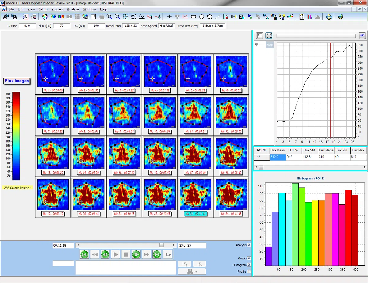
  • High spatial resolution – to catch the finest detail to 100 micron.
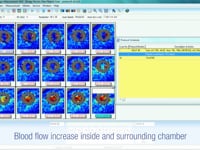
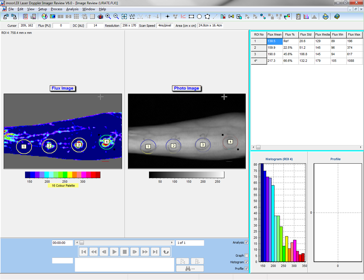
  • Single and Repeat imaging modes – compare flow from region to region within the same scan and scan the same region repeatedly to assess changes over time.
  • Advanced Windows compatible software – to ease setup and scanning. Post Measurement processing functions to make the most of your data.
  • Protocol control – set the imager to control flexible tissue heating, pressure cuff control and transdermal drug delivery routines – reproducible, precise and reliable.
  • Digital Trigger In/ Out – to synchronise with external devices.
  • Digital Signal Processing and High Quality optics – providing the highest sensitivity to changes in blood flow and superb reliability.
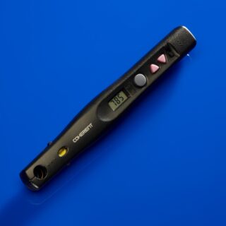
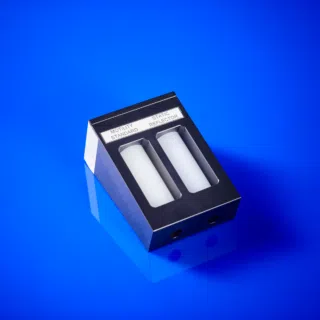
  • Choice of lasers – to assess from the surface to superficial and deeper tissue beds.
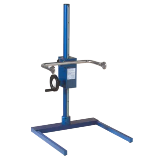
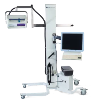
  • Choice of stands – for benchtop and clinical/ theatre use.

For the full range of Moor Instruments imagers, please download the imager catalogue here.

The following products are AVAILABLE TO BUY ONLINE and work with the moorLDI2-IR


This section lists the more common questions our customers have with regards to the moorLDI2-IR. If you have a question you would like answered that does not appear below then please email us. We are happy to help!


Q. What is the largest area you can scan in one image?
A. The moorLDI2-IR will scan a 50x50cm area comfortably. For the highest resolution, choose moorLDI2-HIR - this will scan 2.5cm x 2.5cm with 512 x 512 pixels - around 50 pixels for each square mm!

Q. How often should the moorLDI2 be calibrated?
A. We supply a sealed calibration block to allow the user to easily check and calibrate their system if necessary. We recommend regular checking (weekly) but in practice re-calibration is rarely required.

Q. How can I assess responses to Iontophoresis or Skin Heating with moorLDI2?
A. Set the imager to repeat scan mode so it is scanning the same area repeatedly (you can choose the number of scans and time between each). Clear heating chambers and iontophoresis chambers are available from Moor that allow the imager to 'see' the area that is being stimulated to allow you to compare changes in flow at the stimulated site relative to surrounding tissue.

Q. What are the advantages of a continuously scanning beam?
A. The moorLDI2 uses the patented continuous scanning technique. This means the laser beam does not stop during measurement resulting in a much faster scan. This fast mode is essential for large area clinical assessments. The movement pattern is smooth, deliberate and well defined. For low flow applications we recommend slowing the beam - easily done through scan configuration.

There are numerous references where our laser Doppler imagers are cited. The list below is a small selection. Please contact us for reference lists on your chosen subject.


Abraham, A., Alabdali, M., Alsulaiman, A., Breiner, A., Barnett, C., Katzberg, H.D., Lovblom, L.E., Perkins, B.A., Bril, V., (2016).
Laser Doppler Flare Imaging and Quantitative Thermal Thresholds Testing Performance in Small and Mixed Fiber Neuropathies.
Plos One, 11(11), ppe0165731.

Campos-Junior, P. H. A., Alves, T. J. M., Dias, M. T., Assunçao, C. M., Munk, M., Mattos, M. S., Kraemer, L. R., Almeida, B. G., Russo, R. C., Barcelos, L., Camargo, L. S. A., and Viana, J. H. M., (2016).
Ovarian Grafts 10 Days after Xenotransplantation: Folliculogenesis and Recovery of Viable Oocytes.
PLoS ONE 11(6): e0158109. doi:10.1371/journal.pone.0158109.

Chalothorn, D., Clayton, J .A, Zhang, H., Pomp , D., and Faber, J. E., (2007).
Collateral density, remodeling, and VEGF-A expression differ widely between mouse strains.
Physiological genomics, 30(2), pp.179–91.

Datta , D., Ferrell, W . R., Sturrock , R . D., Jadhav , S . T., and Sattar , N., (2007).
Inflammatory suppression rapidly attenuates microvascular dysfunction in rheumatoid arthritis.
Atherosclerosis, 192(2), pp.391–5.

De, P., Peng , Q., Traktuevc, D . O., Li , W., Yoder, M . C., March, K . L., Durden, D . L. (2009).
Expression of RAC2 in endothelial cells is required for the postnatal neovascular response.
Experimental Cell Research, 315(2), pp.248–263.

Dimitroulas, T., Sandoo, A., Hodson, J., Smith, J. P., and Kitas, G. D., (2016).
In vivo microvascular and macrovascular endothelial function is not associated with circulating dimethylarginines in patients with rheumatoid arthritis: a prospective analysis of the DRACCO cohort.
Scand J Clin Lab Invest, 76(4), pp:331-7.

Ebadi, H., Perkins, B . a, Katzberg, H . D., Lovblom, L . E., Bril, V.,(2012).
Evaluation of proxy tests for SFSN: evidence for mixed small and large fiber dysfunction.
PloS one, 7(8), p.e42208.

Edmunds, MC., Wigmore, S., Kluth, D., (2013).
In situ transverse rectus abdominis myocutaneous flap: a rat model of myocutaneous ischemia reperfusion injury.
J Vis Exp. 2013 Jun 8;(76). www.jove.com/video/50473/in-situ-transverse-rectus-abdominis-myocutaneous-flap-rat-model

Gil, C. H., Ki, B. S., Seo, J., Choi, J. J., Kim, H., Kim, I. G., Jung, A. R., Lee, W-Y., Choi, Y., Park, K., Moon, S-H., and Chung, H-M., (2016).
Directing human embryonic stem cells towards functional endothelial cells easily and without purification.
Tissue Engineering and Regenerative Medicine. 13(3), pp:274–283.

Gorodkin, R., Herrick, A. L., and Murray, A. K., (2016).
Microvascular response in patients with complex regional pain syndrome as measured by laser Doppler imaging.
Microcirculation, 23(5), pp:379-83.

Gunawardena, H., Harris, N . D., Carmichael, C., McHugh, N . J., (2007a).
Maximum blood flow and microvascular regulatory responses in systemic sclerosis.
Rheumatology (Oxford, England), 46(7), pp.1079–82.

Gunawardena, H., Harris, N . D., Carmichael, C., McHugh, N . J., (2007b).
Microvascular responses following digital thermal hyperaemia and iontophoresis measured by laser Doppler imaging in idiopathic inflammatory myopathy.
Rheumatology (Oxford, England), 46(9), pp.1483–6.

Hsieh, M.J., Liu, H.T., Wang, C.N., Huang, H.Y., Lin, Y., Ko, Y.S., Wang, J.S., Chang, V.H., and Pang, J.S., (2016).
Therapeutic potential of pro-angiogenic BPC157 is associated with VEGFR2 activation and up-regulation.
J Mol Med (Berl), Nov 15.[Epub ahead of print].

Isenberg, J . S., Maxhimer , J . B., Powers, P., Tsokos, M ., Frazier, W . a, Roberts , D . D., (2008).
Treatment of liver ischemia-reperfusion injury by limiting thrombospondin-1/CD47 signaling.
Surgery, 144(5), pp.752–61.

Isenberg , J . S., Shiva, S., Gladwin, M., (2009).
Thrombospondin-1-CD47 blockade and exogenous nitrite enhance ischemic tissue survival, blood flow and angiogenesis via coupled NO-cGMP pathway activation.
Nitric oxide : biology and chemistry / official journal of the Nitric Oxide Society, 21(1), pp.52–62.

Khoo, T . L., Halim, a S., Zakaria, Z., Mat Saad, a Z., Wu, L . Y., Lau, H . Y.,(2011).
A prospective, randomised, double-blinded trial to study the efficacy of topical tocotrienol in the prevention of hypertrophic scars. Journal of plastic, reconstructive & aesthetic surgery :
JPRAS, 64(6), pp.e137–45.

Krishnan, S . T., Quattrini, C., Jeziorska M., Malik, R.A., Rayman, G., (2007).
Neurovascular Factors in Wound Healing in the Foot Skin of Type 2 Diabetic Subjects.
DIABETES CARE,, 30(12), pp.3058 – 3062.

Kvietys, P., Granger, D., (2012).
Role of reactive oxygen and nitrogen species in the vascular responses to inflammation.
Free Radical Biology and Medicine, 52(3), pp.556–592.

Jensen, A. R., Manning, M. M., Khaneki, S., Drucker, N. A., and Markel, T. A., (2016).
Harvest Tissue Source Does Not Alter the Protective Power of Stromal Cell Therapy Following Intestinal Ischemia and Reperfusion Injury.
Journal of SurgicalResearch, 204(2), pp:361-370.

Lasch, M., Caballero-Martinez, A., Troidl, K., Schloegl, I., Lautz, T., and Deindl, E., (2016).
Arginase inhibition attenuates arteriogenesis and interferes with M2 macrophage accumulation.
Lab Invest. 2016 May 30, doi: 10.1038/labinvest.2016.62. [Epub ahead of print].

Lee, J. H., Ryu, J. M., Han, Y. S., Zia, M. F., Kwon, H. Y., Noh, H., Han, H. J., and Lee, S. H., (2016).
Fucoidan improves bioactivity and vasculogenic potential of mesenchymal stem cells in murine hind limb ischemia associated with chronic kidney disease.
J Mol Cell Cardiol, 97, pp:169-17.

Lohman , B., (2007).
The effect of whole body vibration on lower extremity skin blood flow in normal subjects.
Med Sci …, 13(2).

Marks, D. J. B., Harbord, M . W. N., Macallister, R., Rahman, F . Z., Young , J., Al-lazikani, B., Lees, W., Novelli, M., Bloom, S., Segal, A . W., (2006).
Defective acute inflammation in Crohn’s disease : a clinical investigation.
Lancet, 367.

Nabavi Nouri, M., Ahmed, A., Bril, V., Orszag, A., Ng, E., Nwe, P., Perkins, B . a, (2012).
Diabetic neuropathy and axon reflex-mediated neurogenic vasodilatation in type 1 diabetes.
PloS one, 7(4), p.e34807.

Oda, M., Toba, K., Ozawa, T., Kato, K., Yanagawa, T., Ikarashi, N., Takayama, T., Suzuki, T., Hanawa, H., Fuse, I., Nakata, K., Narita, M., Takahashi, M., Aizawa, Y., (2010).
Establishment of culturing system for ex-vivo expansion of angiogenic immature erythroid cells, and its application for treatment of patients with chronic severe lower limb ischemia.
Journal of molecular and cellular cardiology, 49(3), pp.347–53.

Pinto, N.C., Cassini-Vieira, P., de Souza-Fagundes, E.M., Barcelos, L.S., Nogueira Castañon, M.C., and Scio, E., 2016.
Pereskia aculeata Miller leaves accelerate excisional wound healing in mice.
J Ethnopharmacol., Sep 3. [Epub ahead of print].

Silva, L.P., Pirraco, R.P., Santos, T.C., Novoa-Carballal, R., Cerqueira, M.T.,2, Reis R.L., Correlo, V.M., and Marques, A.P., (2016).
Neovascularization Induced by the Hyaluronic Acid-Based Spongy-Like Hydrogels Degradation Products.
ACS Appl Mater Interfaces, 8(49). pp33464-33474.

Terkelsen, A. J., Bach, F. W., Jensen, T . S., (2009).
Experimental forearm immobilization in humans reduces capsaicin-induced pain and flare.
Brain research, 1263, pp.43–9.

Tsatralis, T., Ridiandries, A., Robertson, S., Vanags, L.Z., Lam, Y.T., Tan, J.T.M., Ng, M.K.C., and Bursill, C.A., 2016.
Reconstituted high-density lipoproteins promote wound repair and blood flow recovery in response to ischemia in aged mice.
Lipids in Health and Disease . 15(1), pp150-161.

Vasam, G., Joshi, S., Jarajapu, Y. P. R., (2016).
Impaired Mobilization of Vascular Reparative Bone Marrow Cells in Streptozotocin-Induced Diabetes but not in Leptin Receptor-Deficient db/db Mice.
Scientific Reports 6(26131), pp:1-13.

Wajima, D., Nakamura, M., Horiuchi, K., Miyake, H., Takeshima, Y., Tamura, K., Motoyama, Y., Konishi, N., Nakase, H., (2010).
Enhanced cerebral ischemic lesions after two-vein occlusion in diabetic rats.
Brain research, 1309, pp.126–35.

Wester, T., Häggblad, E., (2011).
Assessments of skin and tongue microcirculation reveals major changes in porcine sepsis.
Clinical Physiology

Winchester, L., Chou,N ., (2008).
Measurement of sublingual blood velocity as a tool for monitoring sepsis.
Engineering in Medicine and …, (1), pp.3739–3742.

Yang, Y., Chen, Z., Zhang, T., Wang, S., and Qing, Y., (2016).
Wnt3a promoted the therapeutic of angiogenesis on lower leg ischemia with endothelial progenitor cells.
Int J Clin Exp Med, 9(3), pp:5902-5911.

Zeng, L., Xiao, Q., Chen, M., Margariti, A., Martin, D., Ivetic, A., Xu, H., Mason, J., Wang, W., Cockerill, G., Mori, K., Li, J . Y., Chien, S., Hu, Y., Xu, Q., (2013).
Vascular endothelial cell growth-activated XBP1 splicing in endothelial cells is crucial for angiogenesis.
Circulation, 127(16), pp.1712–22.

Zhao, Y., Li, Y., Luo, P., Gao, Y., Yang, J., Lao, K-H., Wang, G., Cockerill, G., Hu, Y., Xu, Q., Li, T., and Zeng, L., (2016).
XBP1 splicing triggers miR-150 transfer from smooth muscle cells to endothelial cells via extracellular vesicles.
Sci. Rep. 6, 28627.

Moor Instruments are committed to product development. We reserve the right to change the specifications below without notice.

The moorLDI2-IR is a Class IIa Active device for diagnosis under EC directive 93/42/EEC 14th June 1993 Medical Device Directive.


LASER SOURCE

Infra-Red Laser Diode: 785nm nominal, maximum power 2.5mW
Ocular Hazard Distance 20m.
Class 3R per IEC 60825-1:2014 and complies with FDA performance standards for laser products except for deviations pursuant to Laser Notice No. 50, dated June 24, 2007
Visible Laser Diode (target beam for infrared systems): 660nm nominal, maximum power 0.25mW
All measurements include cumulative measurement uncertainties and expected increases after manufacture.

PROTECTIVE EYEWEAR REQUIREMENTS

The nominal ocular hazard distance is 20 metres.
Operator protection: OD4, 770-850nm.
Patient protection: OD4, 630-670nm and 770-850nm.

ENVIRONMENT CONDITIONS

Temperature: 15°C to 30°C
Humidity: 20% to 80%
Atmospheric pressure: within the range 86.0 kPa to 106.0 kPa (645mmHg to 795mmHg). Flammable Anaesthetics: the system must not be operated in the presence of flammable anaesthetics.

BANDWIDTH

Scan rate dependent: low frequency cut-off (3db) 20Hz, 100Hz or 250Hz.
Selectable upper cut-off frequency (0.1db) 3KHz, 15KHz or 22.5KHz.

Default Bandwidth in Bold.

RANGE AND SCAN AREA

At 20cm distance, Normal area = 6.6cm x 6.6cm; Large Area =13cm x 13cm.
At 100cm distance, Normal Area = 25cm x 25cm; Large Area = 50cm x 50cm.

SCAN SPEED

Scan speed is approximately 4ms/pixel, 10ms/pixel or 50ms/pixel (at maximum resolution).
Scan duration is typically 40 seconds for a 12.5cm x 12.5cm image at 64 x 64 pixel resolution, about 6 minutes for a 50cm x 50cm image at 256 x 256 pixel resolution at 4ms/pixel and 100cm distance.

SPATIAL RESOLUTION

Up to 512 x 512 pixels (actual measurements not by interpolation): 0.1mm/pixel at 20cm, ‘normal scan’; 1.0mm/pixel at 100cm, ‘large scan’.

LIGHTING CONDITION

Normal, ambient room lighting.

MEASUREMENTS

FLUX Accuracy: ± 10% relative to Moor Instruments moorLDI2 standard’
Precision: ± 3% of measurement value
Range: 0-5000PU

CONC Accuracy: ± 10%
Precision: ± 5% of measurement value
Range: 0-5000AU

DC Accuracy: ± 10%
Precision: ± 3%
Range: 0-5000AU

CMOS CAMERA

Colour, Auto Focus, 2592 x 1944 pixel resolution, 1296 x 972 (2 x binned) pixel resolution.

SOFTWARE

Windows based control, processing and analysis.

ELECTRICAL SAFETY CLASSIFICATION

Type of protection against electric shock – Class I.
Degree of protection against electric shock – Non-patient contact, no applied part.
Degree of protection against ingress of liquid – IPXO (not protected).
Degree of protection against flammable anaesthetics – equipment not suitable for use in the presence of flammable anaesthetics.
Mode of operation – continuous

GENERAL

Universal voltage switch mode power supply, 100-230V, 50-60Hz, 50VA power consumption
Scan Head: Dimensions W H D mm 426 x 244 x 300: Weight 9kgs.
Operating temperature: 15-30°C.

STORAGE AND TRANSPORTATION CONDITIONS

Temperature: 0-45°C.
Humidity: 0-80% RH.
Atmospheric Pressure: 50.0-106.0 kPa.