Over the last few weeks, in the run up to the World Congress of Microcirculation, Beijing in September 2023, we have covered several facets of one of our key applications, hind limb ischemia, in a series of articles. In this, the fourth and final article in the series, we focus on the physiology of ischemia reperfusion in relation to biomarkers.
The series includes Hind Limb Ischemia Techniques, Cell Treatments, Drug Therapies & Treatment Devices and Physiological studies.
All of the papers in this series of short overviews contribute to an understanding of the physiology of hind limb ischemia reperfusion.
There are close to 1000 studies referred to within the publications. These provide a wealth of information well beyond the topics mentioned here. The moorLDI and moorFLPI blood flow imagers continue to contribute to studies using the hind limb ischemia model, providing useful functional outcome measures with physiological and clinical importance.
Video Presentation available now… We hope the series has been interesting and informative and are excited to announce a video overview touching on some of the topics. Please click here to view in our theory section.
Hind limb ischemia and Recent Studies on the Physiology of Ischemia Reperfusion: Biomarkers
In this overview are studies on the physiological changes following hind limb ischemia without therapeutic intervention. Multiple assays are performed in addition to LDI or LSI; their interpretation is beyond the scope of this overview so the biomarkers are simply listed:
Scalabrin et al studied the temporal remodelling of the gastrocnemius muscle with changes in the molecular catabolic-autophagy signalling network, biomarkers studied included: protein content of LC3-I and LC3-II, mitochondrial (Drp1, OPA1, and Mfn2); Sestrin1 and Sestrin2 and expression of the cytosolic antioxidant superoxide dismutase 1 and 2; AMP-activated protein kinase (AMPK) phosphorylation; and antioxidant enzyme expression.
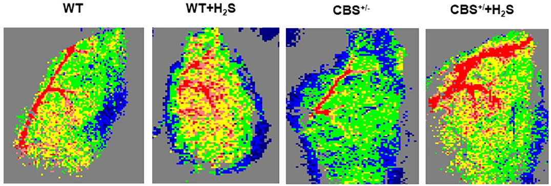
Singh et al investigated whether hydrogen sulphide mitigates skeletal muscle mitophagy-led tissue remodelling via epigenetic regulation of the gene writer and eraser function; biomarkers included: homocysteine; DNA methyltransferase; FTO and TET2; H3K9; GADD45, and MMP-2 & 13.
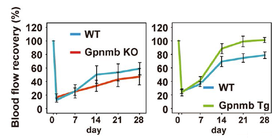
Suda et al investigated the regulation of lysosomal integrity and lifespan of senescent cells by glycoprotein nonmetastatic melanoma protein B. Biomarkers included: short hairpin RNA targeting GPNMB; GPNMB-mCherry protein; accumulation of dysfunctional lysosomes; co-localized with the mitochondria; V-type ATPase; MITF/TFE transcription factors. HLI tests were applied to Gpnmb KO and Gpnmb Transgenic mice and littermate controls:
Vasam et al investigated the early onset of aging phenotype in vascular repair by Mas receptor deficiency. Biomarkers included: number of circulating LSK cells; and mobilization of LSK cells in response to plerixafor or G-CSF. The responses of these to ischemic injury: NO/ROS imbalance in MasR-deficient cells; mean fluorescence intensity of DAF-FM by SDF or VEGF; cellular and mitochondrial ROS levels by using cell- and mitochondria-specific ROS-sensitive fluorescent dyes Cell-ROX and Mito-Sox.
In drawing to a conclusion, we are proud of the impact Laser Doppler (and laser speckle) imaging of the hind limb ischemia model has had both in the reduction in animal use (leading to improved assessment accuracy) and on the understanding of this pathology. We hope the series of articles has covered material that is new and interesting for you or your colleagues.
We hope the diverse range of subjects has been informative and thought provoking. As always, we would be delighted to advise further on any of the instrumentation you might need for this assessment.
We wish the organisers and all participants of the 12th World Congress of Microcirculation in Beijing, China, a very successful, inspirational event!
Hind limb ischemia Physiology
Scalabrin M, Engman V, Maccannell A, Critchlow A, Roberts LD, Yuldasheva N, and Scott Bowen T.
Temporal analysis of skeletal muscle remodeling post hindlimb ischemia reveals intricate autophagy regulation.
Am J Physiol Cell Physiol. 2022 Dec 1; 323(6): C1601–C1610.
doi.org/10.1152/ajpcell.00174.2022
Singh M, Pushpakumar S, Zheng Y, Homme RP, Smolenkova I, Mokshagundam SPL, Tyagi SC.
Hydrogen sulfide mitigates skeletal muscle mitophagy-led tissue remodeling via epigenetic regulation of the gene writer and eraser function.
Physiological Reports. 2022;10:e15422.
doi.org/10.14814/phy2.15422
Figure 6 from this study is shown.
Suda M, Shimizu I, Katsuumi G, Hsiao CL, Yoshida Y, Matsumoto N, Yoshida Y, Katayama A, Wada J, Seki M, Suzuki Y, Okuda S, Ozaki K, Nakanishi‑Matsui M & Minamino T.
Glycoprotein nonmetastatic melanoma protein B regulates lysosomal integrity and lifespan of senescent cells.
Scientific Reports 2022 Apr 20;12(1):6522.
doi.org/10.1038/s41598-022-10522-3
Extracts from Figure 5c and 5d shown.
Vasam G, S SJ, Miyat SY, Adam H, Jarajapu YP.
Early onset of aging phenotype in vascular repair by Mas receptor deficiency.
GeroScience 2022 Feb;44(1):311-327.
doi.org/10.1007/s11357-021-00473-4