Superficial Tissue Oxygenation Monitor

#moorVMS-OXY

Non invasive, real time superficial tissue oxygenation monitor

  • Laser Doppler Imager is a standard accurate method we now use in our cerebral blood flow and brain perfusion in our laboratory.

    Momoh A. Yakubu, PhD
    Texas Southern University

  • Moor Instruments have consistently provided excellent help and support for my research.

    Kim Gooding, PhD
    University of Exeter Medical School

  • We have found Moor equipment to be extremely dependable and innovative.

    Dean L. Kellogg, Jr., MD, Ph.D
    University of Texas Health Science Center

  • In a nutshell, moorFLPI-2 is the most user-friendly system for studying cerebral blood flow regulation in rodents.

    Chia-Yi (Alex) Kuan, MD, PhD
    Emory University School of Medicine

  • We can't recommend Moor instruments highly enough. The technology is at the cutting edge and the support second to none.

    Paul Sumners, PhD
    London South Bank University

  • It goes without saying that the company's imaging technology itself is superb!

    Gourav Banerjee
    Leeds Beckett University

  • I expect to be using Moor Instrument’s technology for many years to come!

    Faisel Khan, PhD
    Ninewells Hospital & Medical School

  • I cannot rate the company or the staff highly enough.

    Jim House, PhD
    University of Portsmouth

The moorVMS-OXY superficial tissue oxygenation monitor uses a white light spectroscopy method to assess superficial tissue oxygen saturation (SO2), tissue haemoglobin concentration and temperature. The technique offers practical advantages such as simplicity in use, real time measurement and the possibility to measure tissue oxygenation, blood flow and temperature with one probe (skin or needle designs) in conjunction with a moorVMS-LDF laser Doppler system and the moorVMS-LDF1-HP high power laser Doppler system. Unlike other techniques, SO2 monitoring does not require tissue heating for reliable baseline measurements enabling true resting oxygenation status to be assessed, as well as adequacy of oxygen delivery to tissue upon heating.

For more demanding research applications, moorVMS-PC supports simultaneous protocol control of pressure cuff and skin heating modules for reproducible provocations. This advanced software also offers standard and custom reports according to your protocol.

The moorVMS-OXY features;

  • Compatibility with all moorVMS modules enabling you to specify your ideal monitoring and provocation system. Stacking case design offers the same compact footprint for multi-module configurations.
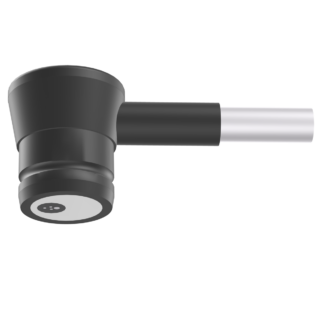
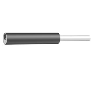
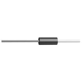
  • Choice of probes – needle and skin designs available. In-built temperature measurement for skin probes. Combined probes offer skin blood flow, tissue oxygenation and temperature measurement in one small probe head. Needle designs combining tissue blood flow and oxygenation measurements.
  • Factory calibrated – no need for complicated setup or probe precalibration.
  • Easy viewing: high contrast, ice white, backlit LCD display.
  • Advanced Windows™ compatible moorVMS-PC software: with extensive automatic analytical features and report generation.
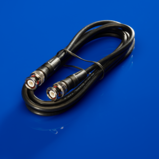
  • Easily connectable: analogue output (0-5V, BNC) and digital (USB) real time data transfer included as standard for connection to data acquisition systems.
  • Medical grade design: for both clinical and research applications.
  • Reliable: 3 year standard warranty, extends to 5 years with annual servicing (in-built automatic reminder).

The following products are AVAILABLE TO BUY ONLINE and work with the moorVMS-OXY


This section lists the more common questions our customers have with regards to the moorVMS-OXY. If you have a question you would like answered that does not appear below then please email us. We are happy to help!


Q. What parameters does the moorVMS-OXY monitor measure?
A. The moorVMS-OXY monitor measures tissue haemoglobin concentration (oxyHb, deoxyHb and totalHb), oxygen saturation (SO2) and temperature.

Q. Why is it important to measure tissue oxygen saturation and total haemoglobin?
A. Tissue haemoglobin and oxygen saturation are two important physiological parameters that indicate tissue health at a microcirculatory level. Oxygen is essential for almost all cells of biological tissue. As well as the continued health of all the cells involved, oxygen is also essential to many aspects of wound healing, including immune function, accumulation of new tissue and growth of new blood vessels. Reduction in oxygen delivery is, therefore, a potentially serious impediment to successful and prompt healing as evidenced by the development of ulcers in patients with peripheral vascular disease. The importance of tissue oxygen measurement in many clinical applications, such as prediction of amputation levels, assessment of reconstructive and plastic surgery, and monitoring of peripheral circulation (e.g. in diabetes patients) have also been recognised for many years.

Q. What is the measurement principle used by moorVMS-OXY?
A. The moorVMS-OXY is based on the theory of white-light reflectance spectroscopy. Its measurement relies on spectrophotometric principles that relate light absorption to chromophore concentrations. The colour spectra of the back-scattered light is analysed to give the overall oxygen saturation of the tissue reflecting and absorbing the light.

Q. Why is haemoglobin concentration unit Arbitrary Unit (AU)?
A. Light scattering confounds the light path in tissue and is generally difficult to quantify. Therefore an absolute quantification of haemoglobin concentrations is difficult. However, the relative concentrations from the oxygenated and deoxygenated haemoglobin and trend measurements are reliable parameters.

Q. How is moorVMS-OXY tissue oxygen saturation different from other measures of oxygenation (SpO2, NIRS, tcpO2)?
A. The moorVMS-OXY SO2 is a measure of the percent haemoglobin oxygen saturation in the capillaries and venous area of the tissue microcirculation and therefore reflects changes in the local conditions of supply and consumption in the tissue. Tissue oxygen saturation is generally lower than arterial oxygen saturation (SaO2) and pulse oximetry saturation (SpO2), and is more close to venous oxygen saturation (SvO2).

SpO2:

SpO2, measured with a pulse oximeter, is the most widely used measure of haemoglobin oxygen saturation. The primary difference is that SpO2 relies on the difference in path length during the pulse cycle to calculate oxygen saturation so that it only measures arterial oxygen saturation. This gives a good indication of lung function, but it gives no information about tissue oxygenation and oxygen uptake by organs as SO2, measured with moorVMS-OXY. In addition, because a pulse oximeter is dependent on detecting the pulse of hemoglobin in the artery, SpO2 requires a pulsatile flow while SO2 readings do not.

NIRS:

Tissue oxygen measurement using near-infrared spectroscopy (NIRS) is also a measure of the local haemoglobin saturation at the measurement site. The difference lies in the light source (white light or laser light), wavelength range (visible region or near infrared region) and algorithms for the derivation of the tissue oxygen saturation. NIRS has been primarily developed to monitor deeper tissues such as muscle and brain while moorVMS-OXY based on the visible light spectroscopy (VLS) measures oxygen saturation in the superficial layer of the tissue. Furthermore, the hemoglobin absorption signal is much stronger in the visible range compared to the NIR, resulting in the higher signal-to-noise ratio and better accuracy with the VLS method.

tcpO2:

tcpO2 is transcutaneous measurement of the oxygen partial pressure. It uses an electrode to heat the underlying tissue to create a local hyperaemia. This means that the transcutaneous measurement values represent the maximum capacity of the vascular bed and tissue to deliver oxygen and transport carbon dioxide away. Therefore, tcpO2 value is close to arterial oxygen saturation and is not a measure of tissue oxygen saturation at normal condition as measured by moorVMS-OXY. The tcpO2 method requires heating the skin tissue to 40°C or higher in order to make a measurement, so it takes several minutes to have the 1st reading and is not suitable for long-term monitoring. In comparison, moorVMS-OXY is a non-invasive, much quick and convenient method for tissue oxygenation measurement. Also, due to the relative large electrode size, tcpO2 cannot be used for measuring oxygen of the internal tissue and in combination with laser Doppler monitor for blood flow measurement at the same site.

Q. What is the measurement depth of moorVMS-OXY?
A. The actual sampling depth depends on probe design and tissue characteristics. In general, the probe with wider separation between delivery and collecting fibres has deeper measurement depth. Physical models predict that the measurements are sensitive to the upper 1 – 2 mm of tissue.

Q. What is the fastest acquisition rate for moorVMS-OXY?
A. The moorVMS-OXY is capable of taking measurements with a rate up to 40 Hz, where heart rate can be clearly visualised in recorded temporal traces of totalHb.

Q. How does skin pigmentation (melanin) affect moorVMS-OXY measurement?
A. As for any device based on reflectance spectroscopy, very strong skin pigmentation might compromise its performance. In such extreme cases, a warning message will be displayed and it is suggested to try alternative measurement sites or set a longer integration time using moorVMS-PC software.

Q. Are there normal / standard values for tissue oxygen saturation?
A. Tissue oxygen saturation is generally lower than arterial oxygen saturation (SaO2) and is more close to venous oxygen saturation (SvO2). Measurement values depend on target tissue site, skin temperature, and patient conditions. Tissue oxygen saturation values for Finger are typically 50% - 80%, and for Leg/Arm are 20% - 50%.

Q. How do I choose which probe to use?
A. The moorVMS-OXY is supplied with a broad probe selection. The standard probes will satisfy most research and clinical purposes. These probes are categorised either by probe type or fibre spacing; Fibre spacing affects penetration depth. Therefore, it is suggested to use skin probe for skin tissues and needle probes for internal tissues. Please consult our sale engineers for more details. We are also happy to discuss any special probe designs or modifications to standard.

Q. Do I need to calibrate the moorVMS-OXY probe before use?
A. No. There is no need to calibrate the probe every time before measurement. The moorVMS-OXY is factory calibrated for each type of probe during manufacture.

Q. Can I regularly check the moorVMS-OXY's performance?
A. Yes. A “Check Probe” is supplied so the user can regularly check the monitor’s integrity.

Q. Does ambient light affect the readings?
A. No. Normally ambient light does not affect the readings because the tissue site is shielded by the probe. To see if the ambient lighting has adverse effect on the measurement, turn the lighting off and on, or cover / uncover the tissue site while reading the SO2 measurements. External lighting that causes a noticeable change in the SO2 and totalHb should be avoided or the tissue shielded from it.

Q. Does probe pressure affect the readings?
A. Yes. High probe pressure will press blood out of the tissue and therefore reduce SO2 and totalHb on skin tissues if not well controlled. All the moorVMS-OXY skin probes are supplied with probe holders and adhesive discs, which will hold the probe on skin firmly with neutral contact pressure.

Q. Is it possible to save the absorption spectra and measurements for post-analysis?
A. Yes. The absorption spectra (raw and reference spectra) and measurements can be continuously saved for post measurement analysis in a text file format using moorVMS-PC software.

Q. Is it possible to measure tissue oxygenation and blood flow simultaneously?
A. Yes. The moorVMS-OXY can be used with the moorVMS-LDF laser Doppler monitor to simultaneously measure SO2, totalHb and blood flow. Combined probe heads are available. The combination of laser Doppler Flux and SO2 measurements would allow clinicians to investigate oxygen metabolism in tissue.

Q. Can I use moorVMS-OXY with other protocol modules?
A. Yes. The moorVMS-OXY is intended to form a modular system when used in combination with the LDF monitor, Skin heater, Iontophoresis controller and Pressure controller. When used in this way the instruments will be controlled and monitored via USB using the moorVMS-PC software.The moorVMS–DAQ is also available to integrate 8 channels of other physiological / analogue data from other systems found in the laboratory or clinic.

Q. Does the technique cause any local heating effects or tissue damage?
A. No. The moorVMS-OXY is a non-invasive device hence causing no damage to target tissues. The light induced heating from the probe tip is negligible. Therefore, moorVMS-OXY is suitable for long term monitoring.

Q. Can the moorVMS-OXY be used for tissues other than skin?
A. Yes. The moorVMS-OXY can also be used for internal tissues, such as brain, with needle probes.

There are numerous references where our tissue oxygenation monitors are cited. The list below is a small selection. Please contact us for reference lists on your chosen subject.


Harrison DK, Evans SD, Abbot NC, Swanson Beck J and McCollum PT.
Spectrophotometric measurements of haemoglobin made saturation and concentration in skin during tuberculin reaction in normal human subjects.
Clin. Phys. Physiol. Meas. 13 349-363. 1992
Weblink

Harrison DK, Habil M.
Optical Measurement of Tissue Oxygen Saturation.
Lower Extremity Wounds 1(3); 191-201. 2002
Weblink

Hongyuan Liu, Matthias Kohl-Bareis, Xiabing Huang
Design of an oxygenation monitor and verification on human skin tissue
European Conferences on Biomedical Optics, Proc. of SPIE Vol. 8087 80871Y-1, 22 - 26 May 2011
Weblink

Kohl M., Lindauer U., Royl G. et al.
Physical Model for the Spectroscopic Analysis of Cortical Intrinsic Optical Signals
Phys Med Biol 45, 3749-3764. 2000
Weblink

Kohl-Bareis, M., Guertler, R., Lindauer, U., Leithner, C., Sellien, H., Royl, G. and Dirnagl, U
System for the Measurement of Blood Flow and Oxygenation in Tissue Applied to Neurovascular Coupling in Brain
Proc. SPIE, 5859. 2005
Weblink

Kuliga KZ, McDonald EF, Gush R, Michel C, Chipperfield AJ, Clough GF.
Dynamics of microvascular blood flow and oxygenation measured simultaneously in human skin.
Microcirculation 2014 Aug;21(6):562-73
Weblink

Sakr, Y.
Techniques to assess tissue oxygenation in the clinical setting
Transfusion and Apheresis Science: Official Journal of the World Apheresis Association: Official Journal of the European Society for Haemapheresis, 43(1), 79-94. 2010
Weblink

Sørensen, H., Rasmussen, P., Siebenmann, C., Zaar, M., Hvidtfeldt, M., Ogoh, S., Sato, Kohei, Kohl-Bareis, M., Secher, N.H., Lundby, C., (2015).
Extra-cerebral oxygenation influence on near-infrared-spectroscopy-determined frontal lobe oxygenation in healthy volunteers: a comparison between INVOS-4100 and NIRO-200NX.
Clinical physiology and functional imaging, 35(3), pp177-184.
Weblink

Sørensen, H., Rasmussen, P., Sato, K., Persson, S., Olesen, N.D., Nielsen, H.B., Olsen, N.V., Ogoh, S., Secher, Niels H., (2014).
External carotid artery flow maintains near infrared spectroscopy-determined frontal lobe oxygenation during ephedrine administration.
British journal of anaesthesia, 113, pp452-8.
Weblink

Sørensen, H., Sc, M., Secher, N. H., Sc, D. M , Siebenmann, C., Rasmussen, P., and Ph, D., (2012).
Cutaneous Vasoconstriction Affects Near-infrared Spectroscopy Determined Cerebral Oxygen Saturation during Administration of Norepinephrine.
American Society of Anesthesiologists., 117(2), pp.263 – 270.
Weblink

Hongyuan Liu, Matthias Kohl-Bareis, and Xiabing Huang (2011).
Design of a tissue oxygenation monitor and verification on human skin.
Proc. SPIE 8087, Clinical and Biomedical Spectroscopy and Imaging II, 80871Y (10 June 2011).
Weblink

Moor Instruments are committed to product development. We reserve the right to change the specifications below without notice.


OPTICS

Light Source White light, wavelength range 400nm – 700nm
Max Output Power 6mW
LED Classification BS EN 62471

MEASURED PARAMETERS

SO2 (tissue oxygen saturation)
Range: 0 – 99%
Accuracy: ± 2 SO2 units
Resolution: 1 SO2 unit

oxyHb (relative oxygenated haemoglobin concentration in arbitrary unit)
Range: 0-1000 AU
Accuracy: ±10%
Resolution: 0.1 AU

deoxyHb (relative deoxygenated haemoglobin concentration in arbitrary unit)
Range: 0-1000 AU
Accuracy: ± 10%
Resolution: 0.1 AU

totalHb (relative total haemoglobin concentration in arbitrary unit)
Range: 0-1000 AU
Accuracy: ± 10%
Resolution: 0.1 AU

Temperature (tissue temperature)
Range: 5 – 50°C
Accuracy: ± 0.3°C
Resolution: 0.1°C

Sampling rate (all parameters) 40Hz

SO2 SIGNAL PROCESSING

Wavelength range: 500 – 650nm
Spectral resolution: 2nm

NUMBER OF MEASUREMENTS

1 x SO2, 1 x oxyHb, 1 x deoxyHb, 1 x totalHb, 1 x Temperature

ANALOGUE OUTPUTS

Range: 0-5V
Resolution: 12-bit
Number of outputs: 4 outputs
Connectors: 50 Ohm BNC sockets

OUTPUTS

Digital LCD screen display

OPERATING ENVIRONMENT

Indoor hospital or laboratory use
Temperature range: 15 – 30°C

GENERAL

Universal voltage switch-mode power supply range 100-230v at 30VA, 50 to 60 Hz
Dimensions W H D mm
moorVMS-OXY 235 x 80 x 200
Weight 1.8kgs
Mode of operation: continuous.

STORAGE AND TRANSPORT CONDITIONS

Temperature 0-45°C
Humidity 0-80% RH
Atmospheric pressure 500-1060hPa
Type of protection against electric shock – Class 1
Degree of protection against electric shock – Type BF
Degree of protection against ingress of liquid – IPXO (not protected)
Degree of protection against flammable anaesthetics – equipment not suitable for use in the presence of flammable anaesthetics

Moor Instruments manufacture a wide range of probes designed to help you assess flow from almost any tissue. We are more than happy to advise on your particular application but hope too that the following general notes are useful.


Skin probes are available in four formats; OXY only, OXY+temp, OXY+flow and OXY+flow+temp, with two main physical configurations, P1 and P2 type designs.

Skin probes are usually fixed to the skin with a probe holder and double sided adhesive discs, although they can be used in other applications (e.g. bone, visceral measurements) and with other equipment (e.g. Iontophoresis). Please refer to the Accessories and Iontophoresis catalogues.

The standard length for all probes is 2 metres. Longer lengths can be supplied on request (code PXL). The range of probe holders for these (and other) probes is described in the accessories page.

Needle probes are amongst the most versatile designs. They can be used for surface measurements, inserted into tissue or used for single vessel measurements. The compact design also lends itself to measurements in tissues with restricted access. Probes can be fixed in position over tissue with a normal laboratory manipulator by clamping onto the black acetal shank.

The P3 type needle probe is available in OXY only or OXY+flow formats, all built into a needle of approx 1.65mm diameter.

The P7 type needle probe is available in OXY+flow format, built into a needle of approx 3.3mm diameter.