Laser Speckle Contrast Imager

#moorFLPI-2

Full-field, video frame rate blood flow imaging

  • In a nutshell, moorFLPI-2 is the most user-friendly system for studying cerebral blood flow regulation in rodents.

    Chia-Yi (Alex) Kuan, MD, PhD
    Emory University School of Medicine

  • Laser Doppler Imager is a standard accurate method we now use in our cerebral blood flow and brain perfusion in our laboratory.

    Momoh A. Yakubu, PhD
    Texas Southern University

  • I expect to be using Moor Instrument’s technology for many years to come!

    Faisel Khan, PhD
    Ninewells Hospital & Medical School

  • It goes without saying that the company's imaging technology itself is superb!

    Gourav Banerjee
    Leeds Beckett University

  • We can't recommend Moor instruments highly enough. The technology is at the cutting edge and the support second to none.

    Paul Sumners, PhD
    London South Bank University

  • Moor Instruments have consistently provided excellent help and support for my research.

    Kim Gooding, PhD
    University of Exeter Medical School

  • I cannot rate the company or the staff highly enough.

    Jim House, PhD
    University of Portsmouth

  • We have found Moor equipment to be extremely dependable and innovative.

    Dean L. Kellogg, Jr., MD, Ph.D
    University of Texas Health Science Center

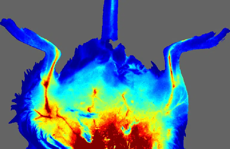
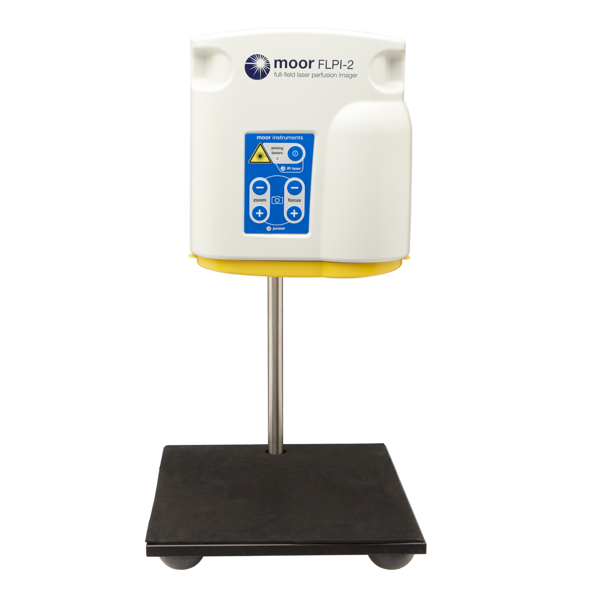
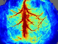
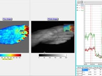
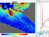
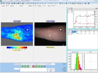
The moorFLPI-2 blood flow imager uses the laser speckle contrast technique to deliver real-time, high-resolution blood flow images, providing outstanding performance in a wide range of pre-clinical and clinical research applications. Optical zoom means you can assess small areas right up to a full size adult hand with a single imager.

Capture every detail with a new unique laser/ single USB camera package. The key specs that matter: pixel resolution of just 3.9 microns (6.6Mpixels/cm2), image sizes ranging from just 6mm x 8mm right up to 225mm x 300mm, using a unique 10x optical zoom function all at up to 100 frames per second…

moorFLPI2 features an ergonomically designed scan head and highly refined software package which promotes a smooth workflow and enables the high through-put required to scan cohorts quickly and accurately. Advanced analysis functions help you to draw sound conclusions from your blood flow images. For full details of the software please click here.

moorFLPI-2 highlights include;

  • Image any exposed tissue (skin or surgically exposed tissues) and species.
  • Easy to use, single USB connection, flexible and simple setup helps get you up and running quickly.
  • Three measurement modes with flexible sampling rates (spatial, temporal and sliding window algorithms) to optimise your data for frame rate, spatial resolution and file size.
  • Best spatial resolution of 3.9 microns per pixel (6.6Mpixels/cm2).
  • Real-time video frames rates – up to 100 frames per second at full field (no windowing).
  • Image areas range from 6mm x 8mm to 225mm x 300mm with a unique, motorised 10x optical zoom and auto focus.
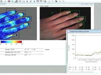
  • Colour photo and blood flow images provided by a unique single USB3 camera, RGB illumination system. Blood flow and photo images precisely matched.
  • Compact design with flexible stand options for laboratory or clinic use.
  • Unique protocol control – automation of pressure cuff, tissue heating and iontophoresis protocols with automated reporting, eases workflow and improves accuracy.
  • CE marking. Well established in the clinical and research communities for best performance and quality.
  • Laser stability hardware for the ultimate in reliable and consistent measurement over minutes, hours and days.

Please contact us, to discuss your specific application requirements with a Moor product specialist. Ask on-line demo to see the new system in action or evaluate it at your own facility.

Please click here for an informative landmark article in the Institute of Physics publication, OLE (Optics and Lasers Europe). The article gives some background to the Full Field technique and offers a comprehensive history, from early beginnings to the current developments at Moor Instruments. Comments are featured from Dr David Briers, the originator of the speckle technique.

The following products are AVAILABLE TO BUY ONLINE and work with the moorFLPI-2


This section lists the more common questions our customers have with regards to the moorFLPI-2. If you have a question you would like answered that does not appear below then please email us. We are happy to help!


Q. How is the moorFLPI-2 connected to the PC?
A. The moorFLPI-2 is connected to the PC via one USB 3 port.

Q. What is the penetration depth of moorFLPI-2?
A. The penetration depth will depend on the optical properties of the tissue sampled as in conventional laser Doppler imaging; however, the signal processed which results in the speckle contrast image is biased towards light coming from the more superficial layers of the tissue. The effective penetration depth will be less than a conventional laser Doppler imager so will focus far more on the nutritive layers and will be less influenced by flow from deeper and larger vessels.

Q. What is the largest area you can image with moorFLPI-2?
A. The image area is dependent on the optical zoom used and the distance from the scan head to the tissue. This allows full flexibility to study areas from 5.6mm x 7.5mm up to 225mm x 300mm (continuously variable with zoom lens).

Q. What is the spatial resolution of the moorFLPI-2?
A. The moorFLPI-2 has a spatial resolution of 3.9 microns per pixel (6.6Mpixels/cm2)

Q. How can I assess dynamic responses with moorFLPI-2?
A. The moorFLPI-2 is used in the same way as a conventional laser Doppler imager - changes from a baseline are commonly assessed. The only difference is in the image acquisition rate with moorFLPI-2 providing 100 images per second opening up many new and exciting research opportunities.

Q. Can I image blood vessels with moorFLPI-2?
A. Yes, if the blood vessels lie in the surface of the tissue being imaged and if the tissue covering the vessels is effectively transparent to the laser light. This makes the technique ideally suited to cerebral imaging and general open surgery

Q. How often do I need to calibrate the system?
A. The moorFLPI-2 is factory calibrated. The user can check zeroing and calibration as frequently as required; we recommend to check calibration monthly.

Q. Can I analyse the data from moorFLPI-2?
A. Images can be analysed in much the same way as a conventional laser Doppler images and sequences allowing comparisons of flow within the same image or in the same subject over time. The video capability also allows the user to define up to 16 regions of interest (that can be varied in size, shape and position) and plot flow in real time from those regions - akin to a 16 channel laser Doppler monitor. Frame grabbing allows the user to select images from the video at flexible or pre defined intervals to build a sequence for analysis.

There are numerous references where our speckle contrast imagers are cited. The list below is a small selection.

Clinical Research
Pre-Clinical Research
Surgical Research

Please contact us for reference lists on your chosen subject.


Clinical Research


Andersen, H. H., Gazerani, P., and Arendt-Nielsen, L., (2016).
High-concentration L-menthol exhibits counter-irritancy to neurogenic inflammation, thermal and mechanical hyperalgesia caused by TRPA1-agonist trans-cinnamaldehyde.
J Pain. pii: S1526-5900(16), pp30065-7.
Weblink

Andersen, H. H., Lundgaard, A. C., Petersen, A. S., Hauberg, L. E., Sharma, N., Hansen, S. D., Elberling, J., and Arendt-Nielsen, L., (2016).
The Lancet Weight Determines Wheal Diameter in Response to Skin Prick Testing with Histamine.
PLoS ONE 11(5).
Weblink

Anna E. Stanhewicz, Sara B. Ferguson, Rebecca S. Bruning, Lacy M. Alexander (2014).
Laser-Speckle Contrast Imaging: A Novel Method for Assessment of Cutaneous Blood Flow in Perniosis.
JAMA Dermatol. 2014;150(6):658-660.

Bahadori, S., Immins, T., Wainwright, T. W (2017).
The Effect of Calf Neuromuscular Electrical Stimulation and Intermittent Pneumatic Compression on Thigh Microcirculation.
Microvascular Research Volume 111, May 2017, Pages 37-41.
Weblink

Bezemer, R., Klijn, E., Khalilzada, M., Lima, A., Heger, M., Bommel , J . Van, and Ince, C., (2010).
Validation of near-infrared laser speckle imaging for assessing microvascular ( re ) perfusion.
Microvascular Research, 79(2), pp.139–143.
Weblink

Cowley, K., and Vanoosthuyze, K., (2016).
The biomechanics of blade shaving.
International Journal of Cosmetic Science. 38(Suppl 1), pp:17-2 3
Weblink

Craighead, D. H., and Alexander, L. M., (2016).
Topical menthol increases cutaneous blood flow.
Microvasc Res., 107, pp:39-45.
Weblink

Dusch, M., Schley, M., Rukwied, R., and Schmelz, M., (2007).
Rapid flare development evoked by current frequency-dependent stimulation analyzed by full-field laser perfusion imaging.
Neuroreport, 18(11), pp.1101–5.
Weblink

Gorbach, a. M., Wang, H., Wiedenbeck, B., Liu, W., Smith, P . D., and Elster, E., (2009).
Functional assessment of hand vasculature using infrared and laser speckle imaging.
Proceedings of SPIE, 7169, pp.716919–716919–9.
Weblink

Jack D Wilkinson, Sarah A Leggett, Elizabeth J Marjanovic, Tonia L Moore, John Allen, Marina E Anderson, Jason Britton, Maya H Buch, Francesco Del Galdo, Christopher P Denton, Graham Dinsdale, Bridgett Griffiths, Frances Hall, Kevin Howell, Audrey MacDonald, Neil J McHugh, Joanne B Manning, John D Pauling, Christopher Roberts, Jacqueline A Shipley, Ariane L Herrick, Andrea K Murray (2018).
A Multicenter Study of the Validity and Reliability of Responses to Hand Cold Challenge as Measured by Laser Speckle Contrast Imaging and Thermography: Outcome Measures for Systemic Sclerosis-Related Raynaud's Phenomenon.
Arthritis Rheumatol. 2018 Jun;70(6):903-911.

Mohammed Ashrafi, Yun Xu, Howbeer Muhamadali, Iain White, Maxim Wilkinson, Katherine Hollywood, Mohamed Baguneid, Royston Goodacre, Ardeshir Bayat (2020).
A microbiome and metabolomic signature of phases of cutaneous healing identified by profiling sequential acute wounds of human skin: An exploratory study.
PLoS ONE 15(2).
Weblink

Rubinder Basson , Mohamed Baguneid , Philip Foden , Rawya Al Kredly , and Ardeshir Bayat (2019).
Functional Testing of a Skin Topical Formulation In Vivo: Objective and Quantitative Evaluation in Human Skin Scarring Using a Double-Blind Volunteer Study with Sequential Punch Biopsies.
Advances in Wound Care VOL. 8, NO. 5
Weblink

S M Jansen, D M de Bruin, M I van Berge Henegouwen, P R Bloemen, S D Strackee, D P Veelo, T G van Leeuwen, S S Gisbertz (2018).
Effect of ephedrine on gastric conduit perfusion measured by laser speckle contrast imaging after esophagectomy: a prospective in vivo cohort study.
Diseases of the Esophagus, Volume 31, Issue 10, October 2018, doy031
Weblink

Tao Huang, Li-Jian Yang, Wei-Bo Zhang, Shu-Yong Jia, Yu-Ying Tian, Guan-Jun Wang, Xiang Mu, Lu Wang, and Gerhard Litscher.
Observation of Microvascular Perfusion in the Hegu (LI4) Acupoint Area after Deqi Acupuncture at Quchi (LI11) Acupoint Using Speckle Laser Blood Flow Scanning Technology.
Evidence-Based Complementary and Alternative Medicine, Volume 2012, Article ID 604590.

Themstrup, L., Welzel, J., Ciardo, S., Kaestle, R., Ulrich, M., Holmes, J., Whitehead, R., Sattler, E. C., Kindermann, N., Pellacani, G., and Jemec, G. B., (2016).
Validation of Dynamic optical coherence tomography for non-invasive, in vivo microcirculation imaging of the skin.
Microvascular Research, 107, pp:97-105.

Timme MAJ van Vuuren, Carina Van Zandvoort, Suat Doganci, Ineke Zwiers, Arina J tenCate-Hoek, Ralph LM Kurstjens, Cees HA Wittens (2017).
Prediction of venous wound healing with laser speckle imaging.
Phlebology. 2017 Dec; 32(10): 658–664.


Pre-Clinical Research


Aubdool, A. A., Kodji, X., Abdul-Kader, N., Heads, R., Fernandes, E. S., Bevan, S., and Brain, S. D., (2016).
TRPA1 activation leads to neurogenic vasodilatation: Involvement of reactive oxygen nitrogen species in addition to CGRP and nitric oxide.
Br J Pharmacol. 173(15), pp:2419-33
Weblink

Bahadori, S., Immins, T., Wainwright, T. W (2017).
A Novel Approach to Overcome Movement Artifact When Using a Laser Speckle Contrast Imaging System for Alternating Speeds of Blood Microcirculation.
J. Vis. Exp. (126), e56415
Weblink

Bezemer, R., Legrand, M., Klijn, E., Heger, M., Post, I . C . J . H., van Gulik, T . M., Payen, D., and Ince, C., (2010).
Real-time assessment of renal cortical microvascular perfusion heterogeneities using near-infrared laser speckle imaging.
Optics express, 18(14), pp.15054–61.
Weblink

Crapser, J., Ritzel, R., Verma, R., Venna, V. R., Liu, F., Chauhan, A., Koellhoffer, W.,, Patel, A., Ricker, A., Maas, K., Graf, J., and McCullough, L. D., (2016).
Ischemic stroke induces gut permeability and enhances bacterial translocation leading to sepsis in aged mice.
Aging (Albany NY). 8(5), pp:1049-63. doi: 10.18632/aging.100952
Weblink

Dean P J Kavanagh, Adam B Lokman, Georgiana Neag, Abigail Colley, Neena Kalia (2019).
Imaging the Injured Beating Heart Intravitally and the Vasculoprotection Afforded by Haematopoietic Stem Cells.
Cardiovasc Res. 2019 Nov 1;115(13):1918-1932.
Weblink

Hashimoto, T., Shibata, K., Nobe, K., Hasumi, K., and Honda, K., (2010).
A Novel Embolic Model of Cerebral Infarction and Evaluation of Stachybotrys microspora Triprenyl Phenol-7 (SMTP-7), a Novel Fungal Triprenyl Phenol Metabolite.
Journal of Pharmacological Sciences, 114(1), pp.41–49.
Weblink

Holstein-Rathlou, Henrik, N., Sosnovtseva , O . V, Pavlov, A . N., Cupples, W . a, Sorensen, C . M., and Marsh, D . J., (2011).
Nephron blood flow dynamics measured by laser speckle contrast imaging. American journal of physiology.
Renal physiology, 300(2), pp.F319–29.
Weblink

Jingli Chen, Jun Yang, Ruiyuan Liu, Chenmeng Qiao, Zhiguo Lu, Yuanjie Shi, Zhanming Fan, Zhenzhong Zhang, Xin Zhang (2017).
Dual-targeting Theranostic System With Mimicking Apoptosis to Promote Myocardial Infarction Repair via Modulation of Macrophages.
Theranostics . 2017 Sep 26;7(17):4149-4167.
Weblink

Jonas Hedelund Rønn, Nikolaj Nerup, Rune Broni Strandby, Morten Bo Søndergaard Svendsen, Rikard Ambrus, Lars Bo Svendsen, Michael Patrick Achiam (2019).
Laser Speckle Contrast Imaging and Quantitative Fluorescence Angiography for Perfusion Assessment.
Langenbecks Arch Surg . 2019 Jun;404(4):505-515.
Weblink

Karalyn E McRae, Jessica Pudwell, Nichole Peterson, Graeme N Smith (2019).
Inhaled Carbon Monoxide Increases Vasodilation in the Microvascular Circulation.
Microvasc Res. 2019 May;123:92-98.
Weblink

Klijn, E., Niehof, S., de Jonge, J., Gommers, D., Ince, C., and van Bommel , J.,(2010).
The effect of perfusion pressure on gastric tissue blood flow in an experimental gastric tube model.
Anesthesia and analgesia, 110(2), pp.541–6.
Weblink

Li Jiang, Mengping Jia, Xiangxiang Wei, Jieyu Guo, Shengyu Hao, Aihong Mei, Xiuling Zhi, Xinhong Wang, Qinhan Li, Jiayu Jin, Jianyi Zhang, Shanqun Li, Dan Meng (2020).
Bach1-induced Suppression of Angiogenesis Is Dependent on the BTB Domain.
EBioMedicine. 2020 Jan;51:102617.
Weblink

Moretti, R., Leger, P. L., Besson, V. C., Csaba, Z., Pansiot, J., Di Criscio, L., Gentili, A., Titomanlio, L., Bonnin, P., Baud, O., and Charriaut-Marlangue, C., (2016).
Sildenafil, a cyclic GMP phosphodiesterase inhibitor, induces microglial modulation after focal ischemia in the neonatal mouse brain.
Journal of Neuroinflammation, 13(95), DOI: 10.1186/s12974-016-0560-4.
Weblink

Mottard N, Berkowitz DE, Santhanam L (2019).
Assessing Renal Microvascular Reactivity by Laser Speckle-Contrast Imaging in Angiotensin-II-Treated Mice.
Int J Nephrol Renovasc Dis. 2020; 13: 45–51.
Weblink

Rikard Ambrus, Rune B Strandby, Niels H Secher, Kim Rünitz, Morten B S Svendsen, Lonnie G Petersen, Michael P Achiam, Lars B Svendsen (2016).
Thoracic Epidural Analgesia Reduces Gastric Microcirculation in the Pig.
BMC Anesthesiol. 2016 Oct 6;16(1):86.
Weblink

Scully, C., Mitrou, N., Braam, B., Cupples, W., and Chon, K., (2016).
Detecting Interactions between the Renal Autoregulation Mechanisms in Time and Space.
IEEE Trans Biomed Eng. [Epub ahead of print], DOI: 10.1109/TBME.2016.2569453.
Weblink

Shen, Y. I., Cho, H., Papa, A. E., Burke, J. A., Chan, X. Y., Duh, E. J., and Gerecht, S., (2016).
Engineered human vascularized constructs accelerate diabetic wound healing.
Biomaterials, 102, pp:107-19.
Weblink

Shih, C-C., Hsu, L-P., Liao, M-H., Yang, S-S., Wang, C-Y., Ho, S-T., and Wu, C-C., (2016).
Role of sterile 20/sps1-related proline/alanine-rich kinase in mice with endotoxic shock.
Journal of Medical Sciences, 36(3), pp.101-107.

Sun, Y .Y., Morozov, Y . M., Yang, D., Li, Y., Dunn, R . S., Rakic, P., Chan, P . H., Abe, K., Lindquist, D . M., and Kuan , C .Y., (2014).
Synergy of Combined tPA-Edaravone Therapy in Experimental Thrombotic Stroke.
PloS one, 9(6), p.e98807.

Teng-Fei Xue, Xu Ding, Juan Ji, Hui Yan, Ji-Ye Huang, Xu-Dong Guo, Jin Yang, Xiu-Lan Sun (2017).
PD149163 Induces Hypothermia to Protect Against Brain Injury in Acute Cerebral Ischemic Rats.
J Pharmacol Sci. 2017 Nov;135(3):105-113.

Teresa Mastantuono, Martina Di Maro, Martina Chiurazzi, Laura Battiloro, Espedita Muscariello, Gilda Nasti, Noemy Starita, Antonio Colantuoni, Dominga Lapi (2018).
Rat Pial Microvascular Changes During Cerebral Blood Flow Decrease and Recovery: Effects of Cyanidin Administration.
Front Physiol . 2018 May 15;9:540.
Weblink

Wang, H., Deng, J., Tu, W., Zhang, L., Chen, H., Wu, X., Li, Y., and Sha, H., (2016).
The hematologic effects of low intensity 650 nm laser irradiation on hypercholesterolemia rabbits.
Am J Transl Res., 8(5), pp:2293-300.

Wang, Z., Schuler, B., Vogel, O., Arras, M., and Vogel, J., (2010).
What is the optimal anesthetic protocol for measurements of cerebral autoregulation in spontaneously breathing mice? Experimental brain research. Experimentelle Hirnforschung.
Expérimentation cérébrale, 207(3-4), pp.249–58.

Weijie Xie, Ting Zhu, Xi Dong, Fengwei Nan, Xiangbao Meng, Ping Zhou, Guibo Sun and Xiaobo Sun (2019).
HMGB1-triggered inflammation inhibition of notoginseng leaf triterpenes against cerebral ischemia and reperfusion injury via MAPK and NF-κB signaling pathways.
Biomolecules 2019, 9(10), 512.

Woitzik, J., Hecht, N., Pinczolits, A., Sandow, N., Major, S., Winkler, MK., Weber-Carsten, S., Dohmen, C., Graf,R., Strong, AJ., Dreier, JP., V . P . C . study group., (2013).
Propagation of cortical spreading depolarization in the human cortex after malignant stroke.
Neurology, First published February 27, 2013

Yanni Lv, Wen Liu, Zhaohui Ruan, Zixuan Xu & Longsheng Fu (2019).
Myosin IIA Regulated Tight Junction in Oxygen Glucose-Deprived Brain Endothelial Cells Via Activation of TLR4/PI3K/Akt/JNK1/2/14-3-3ε/NF-κB/MMP9 Signal Transduction Pathway.
Cellular and Molecular Neurobiology volume 39, pages301–319(2019).

Yu-Chang Yeh., Ming-Jiuh Wang., Chih-Peng Lin., Shou-Zen Fan., Jui-Chang Tsai, W .Z . S., (2012).
Enoxaparin sodium prevents intestinal microcirculatory dysfunction in endotoxemic rats.
Critical Care, 2012, 16:R59

Yu-Yo Sun, Chia-Yi Kuan (2015).
A Thrombotic Stroke Model Based On Transient Cerebral Hypoxia-ischemia.
J Vis Exp. 2015 Aug 18;(102):e52978.

Yu-Yo Sun, Yi-Min Kuo, Hong-Ru Chen, Jonah C. Short-Miller, Marchelle R. Smucker, and Chia-Yi Kuan (2020).
A murine photothrombotic stroke model with an increased fibrin content and improved responses to tPA-lytic treatment.
Blood Adv. 2020 Apr 14; 4(7): 1222–1231.

Zhang, M.J., Sansbury, B.E., Hellmann, J., Baker, J.F., Guo, L., Parmer, C.M., Prenner, J.C., Conklin, D.J., Bhatnagar, A., Creager, M.A., and Spite, M., (2016).
Resolvin D2 enhances postischemic revascularization while resolving inflammation.
Circulation, 134(9), pp666-680.


Surgical Research


Annika Rauh, Dominic Henn, Sarah S Nagel, Amir K Bigdeli, Ulrich Kneser, Christoph Hirche (2019).
Continuous Video-Rate Laser Speckle Imaging for Intra- And Postoperative Cutaneous Perfusion Imaging of Free Flaps.
J Reconstr Microsurg . 2019 Sep;35(7):489-498.
Weblink

Hecht, N., Woitzik, J., Dreier, J . P., and Vajkoczy, P., (2009).
Intraoperative monitoring of cerebral blood flow by laser speckle contrast analysis.
Neurosurgical focus, 27(4), p.E11.
Weblink

Hecht. N, Woitzik. J, König. S, Horn. P , V . P., (2013).
Laser speckle imaging allows real-time intraoperative blood flow assessment during neurosurgical procedures.
Journal of Cerebral Blood Flow & Metabolism, doi:10.103(33), pp.1000–1007.
Weblink

Katharine E. Caldwell, Ross M. Clark, Brittany B. Coffman, Jacquelyn S. Brandenburg, Thomas R. Howdieshell (2018).
Investigation of Open Abdomen Visceral Skin Graft Revascularization and Separation from Peritoneal Contents.
Surgical Science 09(01):24-43.
Weblink

Klijn, E., Hulscher, H . C., Balvers, R . K., Holland, W . P . J., Bakker, J., Vincent , A . J . P . E., Dirven, C . M . F., and Ince, C., (2013).
Laser speckle imaging identification of increases in cortical microcirculatory blood flow induced by motor activity during awake craniotomy.
Journal of neurosurgery, 118(2), pp.280–6.
Weblink

McGuire, P . G., and Howdieshell, T . R., (2010).
The importance of engraftment in flap revascularization: confirmation by laser speckle perfusion imaging.
The Journal of surgical research, 164(1), pp.e201–12.
Weblink

Milstein, D. M. J., Ince, C., Gisbertz, S. S., Boateng, K. B., Geerts, B. F., Hollmann, M. W., Henegouwen, M. I. van B., and Veelo, D. P., (2016).
Laser speckle contrast imaging identifies ischemic areas on gastric tube reconstructions following esophagectomy.
Medicine (Baltimore), 95(25).
Weblink

Muhammad S Hussain, Faisel Khan and Sami Shimi (2019).
Assessment of microvascular perfusion of the stomach in oesophageal surgery using full field laser perfusion imaging - Preliminary Study.
J Surg Practice. 2019;2(1):7.
Weblink

P A Brennan, M T Brands, R Gush, P Alam (2018).
Laser-speckle Imaging to Measure Tissue Perfusion in Free Flaps in Oral and Maxillofacial Surgery: A Potentially Exciting and Easy to Use Monitoring Method.
Br J Oral Maxillofac Surg . 2018 Jul;56(6):556-558.
Weblink

Rikard Ambrus, Michael P Achiam, Niels H Secher, Morten B S Svendsen, Kim Rünitz, Mette Siemsen, Lars B Svendsen (2017).
Evaluation of Gastric Microcirculation by Laser Speckle Contrast Imaging During Esophagectomy.
J Am Coll Surg . 2017 Sep;225(3):395-402.
Weblink

Sanne M Jansen, Daniel M de Bruin, Dirk J Faber, Iwan J G G Dobbe, Erik Heeg, Dan M J Milstein, Simon D Strackee, Ton G van Leeuwen (2017).
Applicability of Quantitative Optical Imaging Techniques for Intraoperative Perfusion Diagnostics: A Comparison of Laser Speckle Contrast Imaging, Sidestream Dark-Field Microscopy, and Optical Coherence Tomography.
J Biomed Opt . 2017 Aug;22(8):1-9.
Weblink

To, Cynthia; Rees-Lee, Jacqueline E.; Gush, Rodney J.; Gooding, Kim M.; Cawrse, Nicholas; Shore, Angela C.; Wilson, Andrew D. (2019).
Intraoperative Tissue Perfusion Measurement by Laser Speckle Imaging: A Potential Aid for Reducing Postoperative Complications in Free Flap Breast Reconstruction.
Plastic and Reconstructive Surgery: 2019 Feb;143(2):287e-292e.


Moor Instruments are committed to product development. We reserve the right to change the specifications below without notice.


Measurement Principle

Laser speckle contrast analysis (also known as LASCA).

Laser Safety Classification

Class 1 per IEC 60825-1:2014 – Safe to use without eye protection.

Calibration

Factory Calibrated.

Image size

from 5.6mm x 7.5mm up to 225mm x 300mm (continuously variable with zoom lens).

Camera/Image Resolution

2064 x 1544 maximum.

Image Acquisition Rate

100 images per second to 1 image every 12 hours.

Acquisition Modes

Single Point (16 channel), Single Image and Video mode.

Optical Design

Motorised 10 x optical zoom and auto focus. Single camera / RGB illumination to match colour photo and blood fl ow images.

Measurement Algorithms

Temporal and Spatial processing (including fixed and sliding window algorithms).

Pixel Resolution

Highest resolution of 6,600,000 pixels per cm².

Software

Refined over 20 years according to customer demands including advanced image acquisition, processing, editing, functionality and analysis.

Stand Options

Scan head has standard VESA mount for desktop stand, Microstand and Clinical Mobile stand. Photographic fitting for use with tripods, etc.

PC Connections

1 x USB 3.0 port.

External Connections

Programmable trigger in / out function with BNC connections.

Warranty

2 years, parts & labour, enhanced service contracts available.

Weights/ Dimensions

Scan head 23cm x 12cm x 25cm, Scan head 2.3kg.

Power Supply

Universal Voltage, 100V-230V. Note acquisition rate is unaffected by frequency of local electrical supply.