High Resolution Laser Doppler Imager

#moorLDI2-HIR

High resolution laser Doppler imager with best spatial resolution of 50 microns

  • In a nutshell, moorFLPI-2 is the most user-friendly system for studying cerebral blood flow regulation in rodents.

    Chia-Yi (Alex) Kuan, MD, PhD
    Emory University School of Medicine

  • I cannot rate the company or the staff highly enough.

    Jim House, PhD
    University of Portsmouth

  • I expect to be using Moor Instrument’s technology for many years to come!

    Faisel Khan, PhD
    Ninewells Hospital & Medical School

  • Laser Doppler Imager is a standard accurate method we now use in our cerebral blood flow and brain perfusion in our laboratory.

    Momoh A. Yakubu, PhD
    Texas Southern University

  • Moor Instruments have consistently provided excellent help and support for my research.

    Kim Gooding, PhD
    University of Exeter Medical School

  • We have found Moor equipment to be extremely dependable and innovative.

    Dean L. Kellogg, Jr., MD, Ph.D
    University of Texas Health Science Center

  • We can't recommend Moor instruments highly enough. The technology is at the cutting edge and the support second to none.

    Paul Sumners, PhD
    London South Bank University

  • It goes without saying that the company's imaging technology itself is superb!

    Gourav Banerjee
    Leeds Beckett University

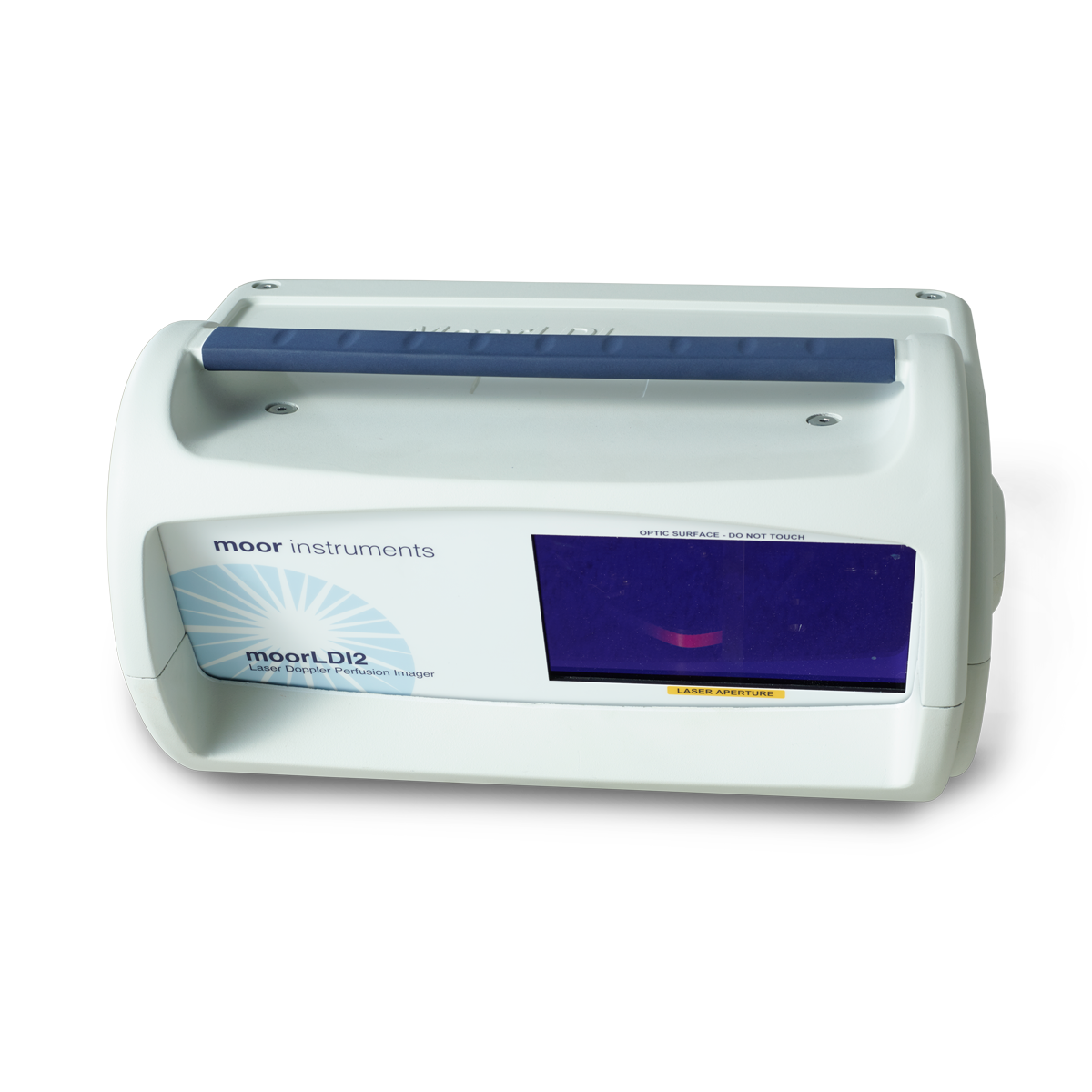
The moorLDI2-HIR high resolution laser Doppler imager is suitable for a wide range of pre-clinical research investigations, more typically where smaller areas are under investigation. The system features unique focused optics to provide 50 micron pixel size and 512 x 512 pixel resolution for high resolution blood flow images. The scan areas range from just 2.5cm x 2.5cm up to 25cm x 25cm with scan times typically less than 5 minutes. Use of a focussed laser provides a deeper measurement depth, optimal for angiogenesis studies such as hind limb ischemia and tumour modelling and pre-clinical cerebral blood flow imaging. Highly refined image measurement and analysis software allows for flexibility in measurement set up and comprehensive analysis functions. The moorLDI2-HIR features a colour photo image of the scanned area and automatic distance measurement, making the positioning and comparison of images easier.

The system is in routine use in numerous laboratories and clinics globally and employs unique, optical design and signal processing in order to generate the highest resolution and clearest images of its class. Laser Doppler imaging (LDI) is often compared to laser speckle imaging and whilst there are some similarities, both techniques offer unique advantages. LDI (and moorLDI2-HIR in particular) generally offers deeper penetration enabling enhanced visualisation of small vessels below the tissue surface, perfect for pre-clinical studies. For certain applications these features are critical.

Other features and benefits include;

  • Non contact measurement – painless for patient, aids infection control, no chemical tracers or dyes needed.
  • Daylight operation – use in most lab, clinic or theatre settings.
  • Flexible scan sizes – from just 2.5cm x 2.5 cm up to 25cm x 25cm.
  • High spatial resolution – to catch the finest detail to 50 micron.
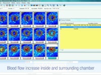
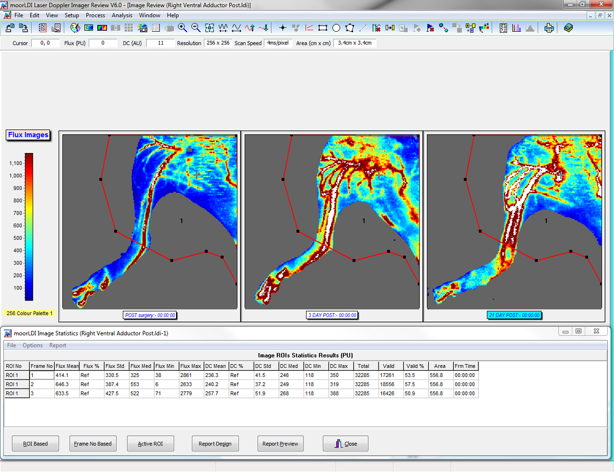
  • Single and Repeat imaging modes – compare flow from region to region within the same scan and scan the same region repeatedly to assess changes over time.
  • Advanced Windows compatible software – to ease setup and scanning. Post Measurement processing functions to make the most of your data.
  • Protocol control – set the imager to control flexible tissue heating, pressure cuff control and transdermal drug delivery routines – reproducible, precise and reliable.
  • Digital Trigger In/ Out – to synchronise with external devices.
  • Digital Signal Processing and high quality optics – providing the highest sensitivity to changes in blood flow and superb reliability.
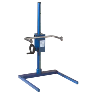
  • Choice of stands – for benchtop use.

NOTE: If you are interested in clinical research and larger scan areas please consider the moorLDI2-IR large area imager or the moorFLPI-2 laser speckle imager.

The following products are AVAILABLE TO BUY ONLINE and work with the moorLDI2-HIR


This section lists the more common questions our customers have with regards to the moorLDI2-HIR. If you have a question you would like answered that does not appear below then please email us. We are happy to help!


Q. What is the largest area you can scan in one image?
A. The moorLDI2-HIR - this will scan 2.5cm x 2.5cm with 512 x 512 pixels equating to around 50 pixels for each square mm, maximum area possible with this system is 25cm x 25cm.

Q. What if I need higher resolution than 50 micron per pixel?
A. The moorFLPI-2 will give you higher resolution, up to 3.9 microns per pixel, for further information on this system, please click here.

Q. What consumables are required for the measurement?
A. The calibration fluid (MOORLDI-CAL-2PFS) has a shelf life of 12 months and needs to be replaced after this time.

The majority of studies using high resolution laser Doppler have investigated vascular responses with the hind limb ischemia model (ligation of the femoral artery), abbreviated to HLI. The ‘Organ’ category includes Brain, Lung, Skin, and Bone.

HLI Angiogenesis
HLI Atherosclerosis & Inflammation
HLI Cells & Genes
Organ (inc Brain, Lung, Skin & Bone)
Wound Healing & Flap Surgery

Please note that the latest version of moorLDI software offers improved pixel resolution – now 512 x 512 up from 256 x 256.

To find out more, please send us a message.

If you are a current moorLDI2-HIR user and your work is missing or miss-categorised, please send us a copy and / or let us know the most appropriate category.


HLI Angiogenesis


Chalothorn , D., Clayton , J., Zhang , H., Pomp , D., and Faber , J . E., 2007.
Collateral density, remodeling, and VEGF-A expression differ widely between mouse strains.
Physiological genomics, 30(2), pp.179–91.
Weblink

Chalothorn , D., Zhang , H., Clayton , J., Thomas , S., and Faber , J . E., 2005.
Catecholamines augment collateral vessel growth and angiogenesis in hindlimb ischemia.
American journal of physiology. Heart and circulatory physiology, 289(2), pp.947–959.
Weblink

Chang, T-T., Lin, L-Y., Chen, J-W (2019).
Inhibition of macrophage inflammatory protein-1β improves endothelial progenitor cell function and ischemia-induced angiogenesis in diabetes.
Angiogenesis. 22(1):53-65.

Chen, L., Zhang, L., Fang, Z., Li, C., Yang, Y., You, X., Song, M., Coffie, J., Zhang, L., Gao, X., Wang, H., (2018).
Naoxintong restores collateral blood flow in a murine model of hindlimb ischemia through PPARδ-dependent mechanism.
Journal of Ethnopharmacology Volume 227, Pages 121-130.

Cheng, G., Zhang, H., Yang, X., Tzima, E., Ewalt, K.L., Schimmel, P., and Faber, J.E., (2008).
Effect of mini-tyrosyl-tRNA synthetase on ischemic angiogenesis, leukocyte recruitment, and vascular permeability.
Am J Physiol Regul Integr Comp Physiol. 295(4): R1138–R1146.

Clayton, J.A., Chalothorn, D., and Faber, J.E., (2008).
Vascular Endothelial Growth Factor-A Specifies Formation of Native Collaterals and Regulates Collateral Growth in Ischemia.
Circ Res. 103(9): 1027–1036.

England, C.G., Im, H-J., Feng, L., Chen, F., Graves, S.A., Hernandez, R., Orbay, H., Xu, C., Cho, S.Y., Nickles, R.J., Liu, Z., Lee, D.S., Cai, W., (2016).
Re-assessing the enhanced permeability and retention effect in peripheral arterial disease using radiolabeled long circulating nanoparticles.
Biomaterials. 100:101-9.

Haywood, N.J., Slater, T.A., Drozd, M., Warmke, N., Matthews, C., Cordell, P.A., Smith, J., Rainford, J., Cheema, H., Maher, C., Bridge, K.I., Yuldasheva, N.Y., Cubbon, R.M., Kearney, M.T., Wheatcroft, S.B., (2020).
IGFBP-1 in Cardiometabolic Pathophysiology—Insights From Loss-of-Function and Gain-of-Function Studies in Male Mice.
Journal of the Endocrine Society, Volume 4, Issue 1, January 2020, bvz006.

Im, H-J., England, C. G., Feng, L., Graves, S. A., Hernandez, R., Nickles, R. J., Liu, Z., Lee, D. S., Cho, S. Y., and Cai, W. (2016).
Accelerated Blood Clearance Phenomenon Reduces the Passive Targeting of PEGylated Nanoparticles in Peripheral Arterial Disease.
ACS Appl. Mater. Interfaces, 8 (28), pp.17955–17963.
Weblink

Im, H-J., England, C-G., Feng, L., Graves, S.A., Hernandez, R., Nickles, R.J., Liu, Z., Lee, D.S., Cho, S.Y., Cai, W., (2016).
Accelerated Blood Clearance Phenomenon Reduces the Passive Targeting of PEGylated Nanoparticles in Peripheral Arterial Disease.
ACS Appl Mater Interfaces. 8(28):17955-63.

Kant, S., Craige, S.M., Chen, K., Reif, M.M., Learnard, H., Kelly, M., Caliz, A.D., Tran, K-V., Ramo, K., Peters, O.M., Freeman, M., Davis, R.J., Keaney Jr, J.F., (2019).
Neural JNK3 regulates blood flow recovery after hindlimb ischemia in mice via an Egr1/Creb1 axis.
Nat Commun. 10(1):4223.

Kawanishi, H., Ohashi, K., Ogawa, H., Otaka, N., Takikawa, T., Fang, L., Ozaki, Y., Takefuji, M., Murohara, T., Ouchi, N., (2020).
A novel selective PPARα modulator, pemafibrate promotes ischemia-induced revascularization through the eNOS-dependent mechanisms.
PLoS One. 15(6): e0235362.

Lee, C-Y., Lin, S-J., Wu, T-C., (2020).
miR-548j-5p Regulates Angiogenesis in Peripheral Artery Disease.
Research Square. Cardiac & Cardiovascular Systems.

Majumder, A., Singh, M., George, A.K., Behera, J., Tyagi, N., and Tyagi, S.C., (2018).
Hydrogen sulfide improves postischemic neoangiogenesis in the hind limb of cystathionine‐β‐synthase mutant mice via PPAR‐γ/VEGF axis.
Physiol Rep. 6(17): e13858.

Murai, Y., Sasase, T., Tadaki, H., Heitaku, S., Imagawa, N., Yamada, T., Ohta, T., (2020).
Analysis of haemodynamics and angiogenic response to ischaemia in the obese type 2 diabetic model Spontaneously Diabetic Torii Leprfa (SDT fatty) rats.
Clin Exp Pharmacol Physiol. 47(4):583-590.

Okuno, K., Naito, Y., Yasumura, S., Sawada, H., Asakura, M., Masuyama, T., & Ishihara, M., (2019).
Haploinsufficiency of Transferrin Receptor 1 Impairs Angiogenesis with Reduced Mitochondrial Complex I in Mice with Limb Ischemia.
Scientific Reports volume 9, Article number: 13658.

Padgett, M. E., McCord, T. J., McClung, J. M., and Kontos, C. D. , (2016).
Methods for Acute and Subacute Murine Hindlimb Ischemia.
J. Vis. Exp. 112, e54166
Weblink

Rodrigues, L.M., Silva, H., Ferreira, H., Renault, M-A., and Gadeau, A-P., (2018).
Observations on the perfusion recovery of regenerative angiogenesis in an ischemic limb model under hyperoxia.
Physiol Rep. 6(12): e13736.

Ruvinov , E., Leor , J., and Cohen , S., 2010.
The effects of controlled HGF delivery from an affinity-binding alginate biomaterial on angiogenesis and blood perfusion in a hindlimb ischemia model.
Biomaterials, 31(16), pp.4573–82.
Weblink

Saito, I., Hasegawa, T., Ueha, T., Takeda, D., Iwata, E., Arimoto, S., Sakakibara, A., Akashi, M., Sakakibara, S., Sakai, Y., Terashi, H., Komori, T., (2018).
Effect of local application of transcutaneous carbon dioxide on survival of random-pattern skin flaps.
Plast Reconstr Aesthet Surg. 71(11):1644-1651.

Shi, Y., Fan, S., Wang, D., Huyan, T., Chen, J., Chen, J., Su, J., Li, X., Wang, Z., Xie, S., Yun, C., Li, X., Tie, L., (2018).
FOXO1 inhibition potentiates endothelial angiogenic functions in diabetes via suppression of ROCK1/Drp1-mediated mitochondrial fission.
Biochimica et Biophysica Acta (BBA) - Molecular Basis of Disease Volume 1864, Issue 7, July 2018, Pages 2481-2494.

Xiong, Y., Chang, L-L., Tran, B., Dai, T., Zhong, R., Mao, Y-C., & Zhu, Y-Z., (2019).
ZYZ-803, a novel hydrogen sulfide-nitric oxide conjugated donor, promotes angiogenesis via cross-talk between STAT3 and CaMKII.
Acta Pharmacologica Sinica volume 41, pages 218–22

Yu, Z., Cai, Y., Deng, M., Li, D., Wang, X., Zheng, H., Xu, Y., Li, W., Zhang, W., (2018).
Fat extract promotes angiogenesis in a murine model of limb ischemia: a novel cell-free therapeutic strategy.
Stem Cell Res Ther. 8;9(1):294.

Zayed, M.A., Yuan, W., Chalothorn, D., Faber, J.E., and Parise, L.V., (2010).
Tumor growth and angiogenesis is impaired in CIB1 knockout mice.
J Angiogenes Res. 2: 17.

Zheng, J., Chen, M., Ye, C., Sun, X., Jiang, N., Zou, X., Yang, H., Liu, H., (2020).
BuZangTongLuo decoction improved hindlimb ischemia by activating angiogenesis and regulating gut microbiota in diabetic mice.
J Ethnopharmacol. 248:112330.


HLI Atherosclerosis & Inflammation


Chang, T-T., Lin, L-Y., Chen, J-W., (2019).
Inhibition of macrophage inflammatory protein-1β improves endothelial progenitor cell function and ischemia-induced angiogenesis in diabetes.
Angiogenesis volume 22, pages53–65
Weblink

Gong, P-Y., Tian, Y-S., Guo, Y-J., Gu, L-F., Li, J-Y., Qi, J., Yu, B-Y., (2019).
Comparisons of antithrombosis, hematopoietic effects and chemical profiles of dried and rice wine-processed Rehmanniae Radix extracts.
J Ethnopharmacol, volume 231:394-402.
Weblink

Ravindran, D., Cartland, S.P., Bursill, C.A., Kavurma, M.M., (2019).
Broad-spectrum chemokine inhibition blocks inflammation-induced angiogenesis, but preserves ischemia-driven angiogenesis.
FASEB J. 33(12):13423-13434.
Weblink

Yun, S., Hu, R., Schwaemmle, M.E., Scherer, A.N., Zhuang, Z., Koleske, A.J., Pallas, D.C., and Schwartz M.A., (2019).
Integrin α5β1 regulates PP2A complex assembly through PDE4D in atherosclerosis.
J Clin Invest. 129(11):4863–4874
Weblink


HLI Cells & Genes


Besnier, M., Gasparino, S., Vono, R., Sangalli, E., Facoetti, A., Bollati, V., Cantone, L., Zaccagnini, G., Maimone, B., Fuschi, P., Da Silva, D., Schiavulli, M., Aday, S., Caputo, M., Madeddu, P., Emanueli, C., Martelli, F., & Spinetti, G., (2018).
MicroRNA-210 enhances the therapeutic potential of bone marrow-derived circulating proangiogenic cells in the setting of limb ischemia.
Molecular Therapy, 26(7), 1694-1705.
Weblink

Bose, R.J.C., Kim, B.J., Arai, Y., Han, I-B., Moon, J.J., Paulmurugan, R., Park, H., Lee, S.H., (2018).
Bioengineered stem cell membrane functionalized nanocarriers for therapeutic targeting of severe hindlimb ischemia.
Biomaterials Volume 185, Pages 360-370.
Weblink

He, W., Wang, P., Chen, Q., Li, C., (2020).
Exercise enhances mitochondrial fission and mitophagy to improve myopathy following critical limb ischemia in elderly mice via the PGC1a/FNDC5/irisin pathway.
Skeletal Muscle 10, 25.

Lam, Y.T., Lecce, L., Yuen, G.S.C., Wise, S.G., Handelsman, D.J., and Ng, M.K.C., (2018).
Androgen action augments ischemia-induced, bone marrow progenitor cell-mediated vasculogenesis.
Int J Biol Sci. 14(14): 1985–1992.
Weblink

Lee, J.H., Kim, S.W., Ji, S.T., Kim, Y.J., Jang, W.B., Oh, J-W., Kim, J., Yoo, S.Y., Beak, S.H., & Kwon, S-M., (2017).
Engineered M13 Nanofiber Accelerates Ischemic Neovascularization by Enhancing Endothelial Progenitor Cells.
Tissue Engineering and Regenerative Medicine volume 14, pages787–802.
Weblink

Lee, J.H., Yoon, Y.M., Han, Y-S., Jung, S.K., Lee, S.H., (2019).
Melatonin protects mesenchymal stem cells from autophagy-mediated death under ischaemic ER-stress conditions by increasing prion protein expression.
Cell Prolif. 52(2):e12545
Weblink

Lee. N.G., Jeung, I.C., Heo, S.C., Song, J., Kim, W., Hwang, B., Kwon, M-G., Kim, Y-G., Lee, J., Park, J-G., Shin, M-G., Cho, Y-L., Son, M-Y., Bae, K-H., Lee, S-H., Kim, J.H., Min, J-K., (2019).
Ischemia‐induced Netrin‐4 promotes neovascularization through endothelial progenitor cell activation via Unc‐5 Netrin receptor B.
1. FASEB J. 34(1):1231-1246.

Lu, H., Wang, F., Mei, H., Wang, S., and Cheng, L., (2018).
Human Adipose Mesenchymal Stem Cells Show More Efficient Angiogenesis Promotion on Endothelial Colony-Forming Cells than Umbilical Cord and Endometrium.
Stem Cells International Volume 2018, Article ID 7537589.

Lu, W., Chen, X., Si, Y., Hong, S., Shi, Z., and Fu, W., (2017).
Transplantation of Rat Mesenchymal Stem Cells Overexpressing Hypoxia-Inducible Factor 2α Improves Blood Perfusion and Arteriogenesis in a Rat Hindlimb Ischemia Model.
Stem Cells Int. 2017: 3794817.

Lu, X-J., and Wang, H-T., (2017).
Reduced Gja5 expression in arterial endothelial cells impairs arteriogenesis during acute ischemic cardiovascular disease.
Exp Ther Med. 14(5): 4339–4343.

Makarevich, P.I., Boldyreva, M.A., Gluhanyuk, E.V., Efimenko, A.Y., Dergilev, K.V., Shevchenko, E.K., Sharonov, G.V., Gallinger, J.O., Rodina, P.A., Sarkisyan, S.S., Hu, Y-C., Parfyonova, Y.V., (2015).
Enhanced angiogenesis in ischemic skeletal muscle after transplantation of cell sheets from baculovirus-transduced adiposederived stromal cells expressing VEGF165.
Stem Cell Res Ther. 26;6:204.

Makarevich, P., Tsokolaeva, Z., Shevelev, A., Rybalkin, I., Shevchenko, E., Beloglazova, I., Vlasik, T., Tkachuk, V., Parfyonova, Y., (2012).
Combined Transfer of Human VEGF165 and HGF Genes Renders Potent Angiogenic Effect in Ischemic Skeletal Muscle.
PLoS ONE, Volume 7, Issue 6, e38776.
Weblink

Min, Y., Han, S., Ryu, H.A., Kim, S-W., (2018).
Human adipose mesenchymal stemcells overexpressing dual chemotactic gene showed enhanced angiogenic capacity in ischaemic hindlimb model.
Cardiovascular Research, Volume 114, Issue 10, Pages 1400–1409.

Nossent, A.Y., Bastiaansen, A.J.N.M., Peters, E.A.B., de Vries, M.R., Aref, Z., Welten, S.M.J., de Jager, S.C.A., van der Pouw Kraan, T.C.T.M., Quax, P.H.A., (2017).
CCR7-CCL19/CCL21 Axis is Essential for Effective Arteriogenesis in a Murine Model of Hindlimb Ischemia.
J Am Heart Assoc. 8;6(3):e005281.

Ricard, N., Zhang, J., Zhuang, Z.W., and Simons, M., (2020).
Isoform-Specific Roles of ERK1 and ERK2 in Arteriogenesis.
Cells. 9(1): 38.

Ryan, T. E., Schmidt, C. A., Alleman, R. J., Tsang, A. M., , Green, T. D., Neufer, P. D., Brown, D. A., and McClung, J. M., (2016).
Mitochondrial therapy improves limb perfusion and myopathy following hindlimb ischemia.
J Mol Cell Cardiol., 1(97), pp:191-196.
Weblink

Shevchenko , E . K., Makarevich , P . I., Tsokolaeva , Z . I., Boldyreva , M., Sysoeva , V . Y., Tkachuk , V., and Parfyonova , Y . V, 2013.
Transplantation of modified human adipose derived stromal cells expressing VEGF165 results in more efficient angiogenic response in ischemic skeletal muscle.
Journal of translational medicine, 11(1), p.138.
Weblink

Suzuki , H., Shibata , R., Kito , T., Ishii , M., Li , P., Yoshikai , T., Nishio , N., Ito , S., Numaguchi , Y., Yamashita , J . K., Murohara , T., and Isobe , K., 2010.
Therapeutic angiogenesis by transplantation of induced pluripotent stem cell-derived Flk-1 positive cells.
BMC cell biology, 11, p.72.
Weblink

Wang, H-H., Wu, Y-J., Tseng, Y-M., Su, C-H., Hsieh, C-L., Yeh, H-I., (2019).
Mitochondrial fission protein 1 up-regulation ameliorates senescence-related endothelial dysfunction of human endothelial progenitor cells.
Angiogenesis. 22(4):569-582.

Wu, J., Hadoke, P. W. F., Takov, K., Korczak, A., Denvir, M. A., and Smith, L. B., (2016).
Influence of Androgen Receptor in Vascular Cells on Reperfusion following Hindlimb Ischaemia.
PLOS ONE, May 9
Weblink

Yang, Y., Xiao, L., Chen, N., Li, Y., Deng, X., Wang, L., Sun., H., and Wu, J., (2016).
Platelet-derived factor V promotes angiogenesis in a mouse hind limb ischemia model.
Journal of Vascular Surger, Volume 65, Issue 4, Pages 1180-1188.e1
Weblink

Zhang, H., Chalothorn, D., Faber. J.E., (2019).
Collateral Vessels Have Unique Endothelial and Smooth Muscle Cell Phenotypes.
Int J Mol Sci. 20(15):3608.

Zhang, Y., Wang, Y., Shao, L., Pan, X., Liang, C., Liu, B., Zhang, Y., Xie, W., Yan, B., Liu, F., Yu, X-Y., and Li, Y., (2020).
Knockout of beta‐2 microglobulin reduces stem cell‐induced immune rejection and enhances ischaemic hindlimb repair via exosome/miR‐24/Bim pathway.
J Cell Mol Med. 24(1): 695–710.


Organ (inc Brain, Lung, Skin & Bone)


Abplanalp, W., Haberzettl, P., Bhatnagar, A., Conklin, D.J., and O'Toole, T.E., (2019).
Carnosine Supplementation Mitigates the Deleterious Effects of Particulate Matter Exposure in Mice.
J Am Heart Assoc. 8(13): e013041.
Weblink

Drucker, N.A., Jensen, A.R., te Winkel, J.P., and Markel, T.A., (2019).
Hydrogen Sulfide Donor GYY4137 Acts Through Endothelial Nitric Oxide to Protect Intestine in Murine Models of Necrotizing Enterocolitis and Intestinal Ischemia.
J Surg Res. 234: 294–302
Weblink

Fonseca, R.C., Bassi, G.S., Brito, C.C., Rosa, L.B., David, B.A., Araújo, A.M., Nóbrega, N., Diniz, A.B., Jesus, I.C.G., Barcelos, L.S., Fontes, M.A.P., Bonaventura, D., Kanashiro, A., Cunha, T.M., Guatimosim, S., Cardoso, V.N., Fernandes, S.O.A., Menezes, G.B., de Lartigue, G., Oliveira, A.G., (2019).
Vagus nerve regulates the phagocytic and secretory activity of resident macrophages in the liver.
Brain Behav Immun. 81:444-454
Weblink

Gohin, S., Javaheri, B., Hopkinson, M., Pitsillides, A.A., Arnett, T.R., and Chenu, C., (2020).
Applied mechanical loading to mouse hindlimb acutely increases skeletal perfusion and chronically enhanced vascular porosity.
Journal of Applied Physiology. Vol. 128, No. 4.
Weblink

Han, Y-S., Kim, S.M., Lee, J.H., Jung, S.K., Noh, H., Lee, S.H., (2019).
Melatonin protects chronic kidney disease mesenchymal stem cells against senescence via PrPC‐dependent enhancement of the mitochondrial function.
J Pineal Res. 66(1):e12535.
Weblink

Kim, S-W., Ryu, H.A., Lee, Y.S., Jeong, I.S., Kim, S., (2019).
Generation of directly reprogrammed human endothelial cells derived from fibroblast using ultrasound.
Journal of Molecular and Cellular Cardiology 126 118–128.
Weblink

Li, X., Saeidi, N., Villiger, M., Albadawi, H., Jones, J.D., Quinn, K.P., Austin Jr, W.G., Golberg, A., Yarmush, M.L., (2018).
Rejuvenation of aged rat skin with pulsed electric fields.
J Tissue Eng Regen Med. 12(12):2309-2318.
Weblink

Meakin, P.J., Coull, B.M., Tuharska, Z., McCaffery, C., Akoumianakis, I., Antoniades, C., Brown, J., Griffin, K.J., Platt, F., Ozber, C.H., Yuldasheva, N.Y., Makava, N., Skromna, A., Prescott, A., McNeilly, A.D., Siddiqui, M., Palmer, C.N.A., Khan, F., and Ashford, M.L.J., (2020).
Elevated circulating amyloid concentrations in obesity and diabetes promote vascular dysfunction.
J Clin Invest. 130(8):4104–4117.
Weblink

Moore, H.M., Drucker, N.A., Hosfield, B.D., Shelley, W.C., Markel, T.A., (2020).
Sildenafil as a Rescue Agent Following Intestinal Ischemia and Reperfusion Injury.
Journal of Surgical Research, Volume 246, Pages 512-518.
Weblink

Okyere, B., Creasey, M., Lebovitz, Y., and Theus, M.H., (2018).
Temporal remodeling of pial collaterals and functional deficits in a murine model of ischemic stroke.
J Neurosci Methods. 01; 293: 86–96.
Weblink

Taheri, S., Yu, J., Zhu, H., Shi, H., and Kindy, M.S., (2019).
Transhemispheric Diaschisis after Unilateral Focal Cerebral Ischemia Reperfusion: A Longitudinal Voxel-Based Study by MRI.
Transl Neurosci Res Rev 2(1):27-37.
Weblink

Wang, C-J., Wu, Y., Zhang, Q., Yu, K-W., Wang, Y-Y., (2019).
An enriched environment promotes synaptic plasticity and cognitive recovery after permanent middle cerebral artery occlusion in mice.
Neural Regen Res . 2019 Mar;14(3):462-469.
Weblink

Yan, T., Zhang, T., Mua, W., Qi, Y., Guo, S., Hu, N., Zhao, W., Zhang, S., Wang, Q., Shi, L., Liu, L., (2020).
Ionizing radiation induces BH4 deficiency by downregulating GTP-cyclohydrolase 1, a novel target for preventing and treating radiation enteritis.
Biochemical Pharmacology Volume 180, 114102.
Weblink


Wound Healing & Flap Surgery


Edmunds, MC., Wigmore, S., Kluth, D., (2013).
In situ Transverse Rectus Abdominis Myocutaneous Flap: A Rat Model of Myocutaneous Ischemia Reperfusion Injury.
J Vis Exp. 2013 Jun 8;(76)
Weblink

Feng, C-J., Perng C-K., Lin C-H., Tsai C-H., Huang P-H., Ma H., (2020).
Intra-arterial injection of human adipose-derived stem cells improves viability of the random component of axial skin flaps in nude mice.
J Plast Reconstr Aesthet Surg. 73(3):598-607.
Weblink

Ho, T-J., Chen, J-K., Li, T.S., Lin, J-H., Hsu, Y-H., Wu, J-R., Tsai, W-T., & Chen, H-P., (2020).
The curative effects of the traditional Chinese herbal medicine “Jinchuang ointment” on excisional wounds.
Chinese Medicine volume 15, Article number: 41.
Weblink

Masuda, H., Sato, A., Shizuno, T., Yokoyama, K., Suzuki, Y., Tokunaga, M., Asahara, T., (2019).
Batroxobin accelerated tissue repair via neutrophil extracellular trap regulation and defibrinogenation in a murine ischemic hindlimb model
PLoS One. 16;14(8):e0220898.
Weblink

Millenaar, D., Bachmann, P., Böhm, M., Custodis, F., Schirmer, S.H., (2020).
Effects of edoxaban and warfarin on vascular remodeling: Atherosclerotic plaque progression and collateral artery growth.
Vascular Pharmacology - Volume 127, 106661
Weblink

Sönmez , T . T., Vinogradov , A., Zor , F., Kweider , N., Lippross , S., Liehn , E . A., Naziroglu , M., Hölzle , F., Wruck , C., Pufe , T., and Tohidnezhad , M., (2013.).
The effect of platelet rich plasma on angiogenesis in ischemic flaps in VEGFR2-luc mice.
Biomaterials, 34(11), pp.2674–82.
Weblink

Wu, H., Chen, H., Zheng, Z., Li, J., Ding, J., Huang, Z., Jia, C., Shen, Z., Bao, G., Wu, L., Mamun, A.A., Xu, H., Gao, W. & Zhou, K. (2019).
Trehalose promotes the survival of random-pattern skin flaps by TFEB mediated autophagy enhancement.
Cell Death & Disease volume 10, Article number: 483.
Weblink

Xiong, Y., Chen, L., Yan, C., Zhou, W., Endo, Y., Liu, J., Hu, L., Hu, Y., Mi, B., Liu, G., (2020).
Circulating Exosomal miR-20b-5p Inhibition Restores Wnt9b Signaling and Reverses Diabetes-Associated Impaired Wound Healing.
Small. 16(3):e1904044.
Weblink


Moor Instruments are committed to product development. We reserve the right to change the specifications below without notice.

The moorLDI2-HIR is a class IIa device under EC directive 93/42/EEC 14 June 1993 Medical Device Directive.


LASER SOURCE

Infra-Red Laser Diode: 785nm nominal, maximum power 2.5mW
Ocular Hazard Distance 20m.
Class 3R per IEC 60825-1:2014. Complies with FDA performance standards for laser products except for deviations pursuant to Laser Notice No. 50, dated June 24, 2007.
Visible Laser Diode (target beam for infrared systems): 660nm nominal, maximum power 0.25mW
All measurements include cumulative measurement uncertainties and expected increases after manufacture.

PROTECTIVE EYEWEAR REQUIREMENTS

The nominal ocular hazard distance is 20 metres.
Operator protection: OD4, 770-850nm.
Patient protection: OD4, 630-670nm and 770-850nm.

ENVIRONMENT CONDITIONS

Temperature: 15°C to 30°C
Humidity: 20% to 80%
Atmospheric pressure: within the range 86.0 kPa to 106.0 kPa (645mmHg to 795mmHg).
Flammable Anaesthetics: the system must not be operated in the presence of flammable anaesthetics.

BANDWIDTH

Scan rate dependent: low frequency cut-off (3db) 20Hz, 100Hz or 250Hz.
Selectable upper cut-off frequency (0.1db) 3KHz, 15KHz or 22.5KHz.

Default Bandwidth in Bold.

RANGE AND SCAN AREA

At 20cm distance, Normal Area = 2.5cm x 2.5cm; Large Area = 5cm x 5cm
At 30cm distance , Normal Area = 3.4cm x 3.4cm; Large Area = 6.8cm x 6.8cm

SCAN SPEED

Scan speed is approximately 4ms/pixel, 10ms/pixel or 50ms/pixel (at maximum resolution). Scan duration is typically 40 seconds for a 12.5cm x 12.5cm image at 64 x 64 pixel resolution, about 6 minutes for a 50cm x 50cm image at 256 x 256 pixel resolution at 4ms/pixel and 100cm distance.

SPATIAL RESOLUTION

Up to 512 x 512 pixels (actual measurements not by interpolation): 0.05mm/pixel at 20cm for the ’normal’ size scan.

LIGHTING CONDITION

Normal, ambient room lighting.

MEASUREMENTS

FLUX Accuracy: ± 10% relative to Moor Instruments moorLDI2 standard’
Precision: ± 3% of measurement value
Range: 0-5000PU

CONC Accuracy: ± 10%
Precision: ± 5% of measurement value
Range: 0-5000AU

DC Accuracy: ± 10%
Precision: ± 3%
Range: 0-5000AU

CMOS CAMERA

Colour, Auto Focus, 2592 x 1944 pixel resolution, 1296 x 972 (2 x binned) pixel resolution

SOFTWARE

Windows based control, processing and analysis.

ELECTRICAL SAFETY CLASSIFICATION

Type of protection against electric shock – Class I.
Degree of protection against electric shock – Non-patient contact, no applied part.
Degree of protection against ingress of liquid – IPXO (not protected).
Degree of protection against flammable anaesthetics – equipment not suitable for use in the presence of flammable anaesthetics.
Mode of operation – continuous.

GENERAL

Universal voltage switch mode power supply, 100-230V, 50-60Hz, 50VA power consumption
Scan Head: Dimensions W H D mm 426 x 244 x 300: Weight 9kgs.
Operating temperature: 15-30°C.

STORAGE AND TRANSPORTATION CONDITIONS

Temperature: 0-45°C.
Humidity: 0-80% RH.
Atmospheric Pressure: 50.0-106.0 kPa.