Laser Speckle Blood Flow and Tissue Oxygenation Imager

#moorO2Flo-2

Simultaneous tissue perfusion and oxygenation imaging

  • I expect to be using Moor Instrument’s technology for many years to come!

    Faisel Khan, PhD
    Ninewells Hospital & Medical School

  • I cannot rate the company or the staff highly enough.

    Jim House, PhD
    University of Portsmouth

  • We have found Moor equipment to be extremely dependable and innovative.

    Dean L. Kellogg, Jr., MD, Ph.D
    University of Texas Health Science Center

  • We can't recommend Moor instruments highly enough. The technology is at the cutting edge and the support second to none.

    Paul Sumners, PhD
    London South Bank University

  • In a nutshell, moorFLPI-2 is the most user-friendly system for studying cerebral blood flow regulation in rodents.

    Chia-Yi (Alex) Kuan, MD, PhD
    Emory University School of Medicine

  • It goes without saying that the company's imaging technology itself is superb!

    Gourav Banerjee
    Leeds Beckett University

  • Laser Doppler Imager is a standard accurate method we now use in our cerebral blood flow and brain perfusion in our laboratory.

    Momoh A. Yakubu, PhD
    Texas Southern University

  • Moor Instruments have consistently provided excellent help and support for my research.

    Kim Gooding, PhD
    University of Exeter Medical School

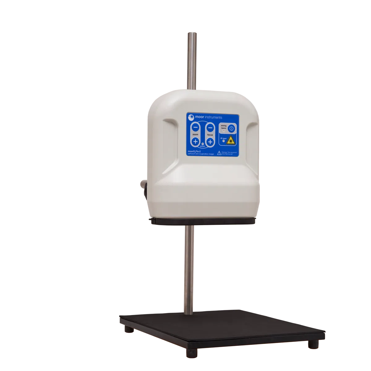
The moorO2Flo-2 perfusion and oxygenation imager is a unique system designed to simultaneously image tissue perfusion and relative tissue oxyhaemoglobin and deoxyhaemoglobin concentration change.

The moorO2Flo-2 combines Moor Instruments’ existing laser speckle contrast technology for perfusion imaging (moorFLPI-2) with reflectance spectroscopy for oxygenation change imaging.

  • Non contact imaging.
  • Blood flow and oxygen videos of any exposed tissue (skin or surgically exposed tissues).
  • Available in Blood flow only mode with imaging at up to 100 frames per second.
  • Combined Blood flow and Oxygen at up to 20 frames per second, with each frame containing 1 x blood flow image, 1 x oxyHb image, 1 x deoxyHB image and 1 x colour photograph.
  • Best spatial resolution of 3.9 microns per pixel to reveal detailed morphology.
  • 10 x optical zoom to image areas from 8mm x 6mm to 300mm x 200mm with motorised zoom and autofocus.
  • Add multiple “regions of interest” to assess and quantify blood flow and oxygen changes both in real time and post measurement. Area of ROIs calculated automatically.
  • Colour photo image matches blood flow and oxygen images precisely to aid identification of features
  • 1 camera – 5 parameters.

**The moorO2Flo-2 is not a medical device. It is intended for use in research and educational life science applications only.**

The following products are AVAILABLE TO BUY ONLINE and work with the moorO2Flo


A. Steimers, M. Gramer, M. Takagaki, R. Graf, U. Lindauer, M. Kohl-Bareis (2013)
Simultaneous Imaging of Cortical Blood Flow and Haemoglobin Concentration with LASCA and RGB Reflectometry
Advances in Experimental Medicine and Biology 789:427-433
Weblink

A. Steimers, M. Gramer, B. Ebert, M. Füchtemeier, G. Royl, C. Leithner, J. P. Dreier, U. Lindauer, M. Kohl-Bareis (2009)
Imaging of cortical haemoglobin concentration with RGB reflectometry
Proceedings Volume 7368, Clinical and Biomedical Spectroscopy; 736813
Weblink

Phill B. Jones; Hwa Kyuong Shin; David A. Boas; Bradley T. Hyman; Michael J. Moskowitz; Cenk Ayata; Andrew K. Dunn (2008)
Simultaneous multispectral reflectance imaging and laser speckle flowmetry of cerebral blood flow and oxygen metabolism in focal cerebral ischemia
Journal of Biomedical Optics, 13(4), 044007
Weblink

Izumi Nishidate, Kiyohiro Sasaoka, Testuya Yuasa, Kyuichi Niizeki, Takaaki Maeda, and Yoshihisa Aizu (2008)
Visualizing of skin chromophore concentrations by use of RGB images
Optics Letters Vol. 33, Issue 19, pp. 2263-2265
Weblink

Elizabeth M. C. Hillman (2007)
Optical brain imaging in vivo: techniques and applications from animal to man
Journal of Biomedical Optics, 12(5), 051402
Weblink

Andrew K. Dunn, Anna Devor, Hayrunnisa Bolay, Mark L. Andermann, Michael A. Moskowitz, Anders M. Dale, and David A. Boas (2003)
Simultaneous imaging of total cerebral hemoglobin concentration, oxygenation, and blood flow during functional activation
Optics Letters Vol. 28, Issue 1, pp. 28-30
Weblink

Ute Lindauer, Georg Royl, Christoph Leithner, Marc Kühl, Lorenz Gold, Jörn Gethmann, Matthias Kohl-Bareis, Arno Villringer, Ulrich Dirnagl (2001)
No Evidence for Early Decrease in Blood Oxygenation in Rat Whisker Cortex in Response to Functional Activation
NeuroImage Volume 13, Issue 6, Pages 988-1001
Weblink

Matthias Kohl, Ute Lindauer, Ulrich Dirnagl, and Arno Villringer (1998)
Separation of changes in light scattering and chromophore concentrations during cortical spreading depression in rats
Optics Letters Vol. 23, Issue 7, pp. 555-557
Weblink

Moor Instruments are committed to product development. We reserve the right to change the specifications below without notice.

WARNING: The moorO2Flo emits flashing light which may potentially trigger seizures for people with photosensitive epilepsy. Do not use the moorO2Flo in the presence of individuals with photosensitive epilepsy.


Quality Control

The moorO2Flo-2 is CE marked.

Measured Parameters

Tissue perfusion, in PU.
Relative oxygenated and deoxygenated haemoglobin concentration change, in AU.

Working Distance

100-380 mm (measured from front edge of enclosure).

Imaging Area

8mm x 6mm (maximum zoom, minimum distance).

300mm x 200mm (minimum zoom, maximum distance).

Maximum image resolution

2060 x 1540 for haemoglobin images

2064 x 1544 for perfusion and colour photo/video images

Maximum frame rate

Up to 20 frames per second for oxygenation imaging or combined perfusion + oxygenation imaging. 1 Frame consists of: 1x perfusion image, 1x oxyHb change image, 1x deoxyHb change image and 1x colour video image.

Up 100 frames per second for perfusion only imaging.

Operating Environment

Indoor laboratory use.
Temperature range: 15 – 30°C.
Atmospheric pressure: 86-106 kPa.
Humidity: 20-80%, non-condensing.
Maximum altitude: 2000 m.
Lighting conditions Oxygenation imaging: near-dark, controlled light environment or used inside a light proof enclosure
Lighting conditions Perfusion only imaging: normal, ambient room lighting

Storage And Transportation Environment

Temperature range: 5 – 45°C.
Atmospheric pressure: 50-106 kPa.
Humidity: 20-80%, non-condensing.

Power Source

AC mains, 100-240 V +/-10%, 0.5A max, 50/60 Hz.

Dimensions

Scan head: 220x112x245 mm (WxHxD), 2.3 kg
PSU: 28x86x56 mm (WxHxD), 0.2 kg

Laser Classification

Class 1 (EN 60825-1:2014+A11:2021).

Measurement Laser

Emission pattern: Diverging circular beam.
Wavelength: 785nm ± 10nm
Maximum power: 90mW, Accessible power below EN60825-1:2014+A11:2021 Class 1 limits.

Aiming Lasers

Emission pattern: Collimated.
Wavelength: 650nm
Maximum power: 0.1mW per laser.

Intended Use

The moorO2Flo-2 is intended for use in research and educational life science applications only. The moorO2Flo-2 is not intended to be used for medical diagnostic or therapeutic purposes.